


The Use of a Second Generation Intramedullary Nail in Fixation of Difficult Ankle and Hindfoot Arthrodeses
Aug 21, 2019George Edward Quill, M.D. (Retired 2023)
Introduction
The goals of ankle and hindfoot arthrodesis are the relief of pain and deformity and the development of a solid fusion. Since the end of the 19th Century, arthrodesis has been an accepted treatment for painful arthritis of the ankle and subtalar joints.1, 3-8, 10-15, 18, 20, 21, 23-29, 31-34 For patients with disabling arthritis, infection, or deformity of the tibiotalar and the talocalcaneal joint, not only must the ankle (tibiotalar) joint be fused, but also the subtalar (talocalcaneal) joint also be fused at the same operative setting. This procedure is termed tibiotalocalcaneal arthrodesis. Fusion of the talus to all the bones articulating with it (distal tibial, calcaneus, tarsonavicular, and cuboid) is termed pantalar arthrodesis (Figure 1).14, 15, 17, 18, 22, 25-28, 30, 33, 34
A medullary nail through the plantar aspect of the foot can afford excellent stability and maintain satisfactory position and alignment when performing these fusion procedures.6,8,14,15,20,25,26 Patients undergoing medullary fixation for tibiotalocalcaneal arthrodesis often need not be immobilized or have their activities restricted as severely as patients undergoing similar procedures employing other methods of fixation. Furthermore, neuropathic patients, even in the presence of an acute Charcot process about the hindfoot and ankle, can be managed quite readily by medullary fixation when undergoing ankle and hindfoot arthrodesis.25, 26
History of Tibiotalocalcaneal Arthrodesis and Ankle Fusion Nail Precedents
Ankle arthrodesis was first reported by Albert in 1882.3 Many reports in the English and German literature describe various techniques and provide clinical results for this procedure.1-3,8-10,17,19,20,31 Fusion of the tibia to the talus and the talus to the os calcis has been performed using posterior extra-articular methods, as described by Staples.29
Intramedullary fixation for tibiotalocalcaneal arthrodesis was described as early as 1906 by Lexer, employing a technique that consisted of placing a boiled corpse bone pin through a hole in the calcaneus, talus and tibia.20 In 1915, Albee used the fibula as a vertical spike to perform an ankle and subtalar fusion.2 In the late 1940’s and mid-1950’s, various other authors described techniques for tibiotalocalcaneal arthrodesis, employing a variety of techniques.1,4,6,7,13,19
In a German text entitled “The Practice of Intramedullary Nailing”, published in the 1960’s, Gerhardt Küntscher described a method of combined arthrodesis of the ankle and subtalar joints.18 He described a technique of closed medullary nailing with a conical nail inserted over a guide pin through the sole of the foot. A 12 x 14 mm. nail was used to achieve an interference fit, and no locking of the nail was done. Patients were kept at bed rest for three weeks and then ambulated in plaster.
Tomeno and Danan had an 80 percent consolidation rate for panarthrodeses reported in 1979 using a variety of fixation techniques. Their infection rate was very high.32 In 1988, Russotti, et al, reported on 21 tibiotalocalcaneal arthrodeses employing Steinmann pins and external fixation. Radiographic union was achieved in 85 percent of these patients with satisfactory results.28
Papa and Myerson published a series of 21 pantalar and tibiotalocalcaneal arthrodeses for osteoarthrosis, achieving an 86 percent fusion rate using a transfibular approach and cannulated screws for fixation. Fusion occurred in their series in under five months, even in neuropathic patients.23, 24
Kile, et al, and others reported as early as 1993 the results of tibiotalocalcaneal fusion using a more modern intramedullary nail and a posterior Achilles splitting approach. These authors initially claimed that the technology involved in using the nail was very involved, quite demanding, and that the long-term results were not yet known.14,15
As early as 1989, this author began a series of tibiotalocalcaneal arthrodesis using an angled intramedullary nail for fixation. This was a closed section nail available in 11, 12 and 13 mm. outer diameters that was initially adapted from its originally intended use in fixation of supracondylar femur fractures.26
In 1996 this author reported the results of his clinical series of tibiotalocalcaneal arthrodesis performed on 40 patients with intramedullary fixation. These 40 patients (25 male and 15 female) with a mean age of 47.6 years (range, 14 to 82 years) underwent this procedure for the diagnoses of osteoarthrosis (25 patients), rheumatoid arthritis (6 patients), pseudarthrosis after failed prior attempts at ankle fusion (3 patients), Charcot arthropathy (4 patients), destructive distal tibial giant cell tumor (1 patient), and rigid talipes equinus, resulting from paraplegia (1 patient). With a mean follow-up interval of 30 months (now averaging over 40 months), a 90 percent union rate has been achieved at a mean time to union of 14 weeks.25
Because of some complications related to that nail, including painful retained or fractured interlocking screws, a 10 percent nonunion rate, difficulty accommodating the 8 degree bow using this nail, and a sometimes less than reliable means of targeting instrumentation and fixation, this author has all but abandoned the use of this first, angled nail in favor of a newer second generation, straight nail.
In developing the second generation ankle fusion nail, many of the drawbacks of the first generation nail have been overcome, and certain advances and a more universally applicable nail developed.
This new ankle arthrodesis nail is ideally suited for almost all tibiotalocalcaneal, pantalar, tibiocalcaneal, and revision ankle fusion indications because it affords a rigid, load-sharing fixation device that incorporates a simple, nail-mounted method of compression across the arthrodesis sites. This second generation nail brings the strength and biocompatibility of titanium alloy in the form of a straight, closed section, cannulated nail design available in outside diameters of 10, 11 and 12 mm. Nail lengths of 15 and 18 cm. are available. Custom sizing and patient-matched implants are available from its manufacturer (Biomet, Inc., Warsaw, IN). The nail is manufactured to accept fully-threaded, 5 mm. titanium locking screws, which are precisely and reproducibly inserted through a nail-mounted targeting device (Figure 2).
The option for compression with a nail-mounted in-line compression device that is quite simple and easy to use is greatly appreciated by the surgeon employing this second generation nail. Transfixation screws are inserted from lateral to medial, but can also be applied from medial to lateral if indicated.
This second generation nail is unique in that it affords even more torsional rigidity and better calcaneal purchase than was available with earlier nail designs, through its transcalcaneal locking screw. This screw is inserted from posterior to anterior using the same nail-mounted targeting device as is employed for the proximal locking screws.
The posterior to anterior screw, which passes through the calcaneus and nail, may also be passed through the transverse tarsal joints to aid in fusing the hindfoot and ankle to the midfoot (Figure 1).
Laboratory biomechanical testing has demonstrated that the insertion of the posterior to anterior fixation screw provides 40 percent more torsional rigidity than other existing first generation nails that incorporate lateral to medial transverse screws alone (data on file with Biomet, Inc., Warsaw, IN).
This article is comprised of the author’s clinical experience in applying this second generation ankle fusion nail to a series of 12 patients undergoing ankle and hindfoot fusion for some very challenging and technically demanding indications and pathoanatomy.
Indications for Ankle and Hindfoot Arthrodesis
The indications for tibiotalocalcaneal and tibiocalcaneal arthrodesis, include avascular necrosis of the talus or failed total ankle replacement arthroplasty with subtalar intrusion. The patient with a failed ankle fusion with an insufficient talar body, as well as those patients with rheumatoid or osteoarthritic involvement of these joints, is also a candidate for tibiotalocalcaneal fusion. Other indications include the sequelae of trauma and the severe deformity of untreated clubfoot and neuromuscular disease. Patients with neuroarthropathy, skeletal defects after tumor reconstruction, pseudarthrosis, as well as fixed and various flail deformities about the hindfoot and ankle, are also candidates for tibiotalocalcaneal and, in certain instances, pantalar arthrodesis. When there is significant instability, subluxation, or arthritis involving not only the ankle and hindfoot, but also the transverse tarsal joints, then coupling the midfoot to the hindfoot by fusing the talonavicular, calcaneocuboid, and calcaneonavicular articulations is indicated (Figure 3).25
Contraindications for Ankle and Hindfoot Arthrodesis
Contraindications for tibiotalocalcaneal and pantalar arthrodesis include a dysvascular extremity or one that has a severe active infection. Tibiotalocalcaneal arthrodesis employing medullary fixation inserted through the sole of the foot will be contraindicated in the patient with an insufficient plantar weight bearing surface for padding. Moderately severe and fixed deformities of the ankle, hindfoot, and distal tibia are relative contraindications for closed nailing and arthrodesis because of the difficulty encountered in obtaining a collinear reduction of the tibia, talus and calcaneus by the closed method.26
Surgical Technique
Patient Positioning: The patient is positioned in the lateral decubitus position on a radiolucent operating table with the affected extremity closest to the ceiling. Great care must be taken to pad the patient’s bony prominences, and an axillary roll is used in the recumbent axilla. The nonoperated extremity is flexed at the hip and knee and placed close to the anterior edge of the operating table so it will be out of the way of a lateral to medial projected fluoroscope. A radiolucent table facilitates the use of intraoperative fluoroscopy to image in all planes. General or spinal anesthesia is usually required, and a thigh tourniquet greatly facilitates the plantar dissection.
Surgical Exposure: An incision for the transfibular approach is made at a level approximately 5 cm. proximal to the ankle joint, extending slightly anteriorly and distally on the sinus tarsi to facilitate exposure of the subtalar joint. Great care is taken to note the course of the neurovascular structures and tendons which are protected. The distal portion of the fibula is resected in a beveled fashion at a level approximately 2 cm. proximal to the tibiotalar joint line. The distal portion of the fibula may be morselized for use as an autogenous bone graft.
The tibiotalar joint is denuded of cartilage in a congruent fashion, removing anterior and posterior osteophytes with a chisel and rongeur. Diseased cartilage is removed down to bleeding subchondral cancellous bone, preserving, if possible, the natural concavity of the distal tibial articular surface and the dorsal convex surface of the talus. If necessary, when performing tibiotalocalcaneal arthrodesis, the tibiotalar joint may be provisionally fixed in place with a large, smooth K-wire, while both sides of the subtalar arthrodesis site are prepared with chisels and rongeurs.
Tibiotalar and subtalar arthrotomies can be used for preparing the joint for arthrodesis, as well as providing a site for bone graft insertion, as indicated. The transfibular approach is also useful in correcting significant preoperative varus deformities or loss of length relative to the fibula due to trauma, infection or neoplasm.
An anterolateral ankle arthrotomy may be used and also may be coupled with an anteromedial ankle arthrotomy to correct deformity present across the ankle joint and to prepare the joint surface by moving what remains of the articular cartilage medially. The best position of arthrodesis is neutral dorsiflexion, no more than 3 to 5 degrees of hindfoot valgus, and external rotation symmetric with the contralateral uninvolved extremity. Appropriate external rotation is achieved when the anteromedial crest of the tibia lies parallel with the second ray of the involved foot.
Entry Site Determination: Following the preparation of the bony surfaces, a 4 cm. longitudinal plantar incision is made anterior to the subcalcaneal fat pad, slightly lateral to the midline. Blunt dissection is taken down to the plantar fascia, which is split longitudinally. The intrinsic muscles are swept to the side, and the neurovascular bundle on the sole of the foot is identified and protected. The correct position for the plantar calcaneal entry site is well anterior to the weight bearing surface of the calcaneal tuberosity at approximately 2 cm. posterior to the articulation of the calcaneus with the transverse tarsal joints. In the coronal plane, the entry sites should line up with the center of the tibial medullary canal. A sharp awl may be used to make the plantar calcaneal opening, after which a 3.2 mm. Steinmann pin is inserted through the calcaneus, talus and into the tibia. Alternatively, a guide wire and cannulated drill may be used for this purpose. The subtalar and tibiotalar articular surfaces are reamed with the cannulated drill. This allows access through the calcaneus and talus to the medullary canal of the tibia. It is helpful to have an assistant hold the foot in the appropriate alignment during the trans-medullary reaming.
Guide Wire Placement and Canal Reaming: A ball-tipped guide wire is inserted trans-calcaneally through the talus into the tibial medullary canal using image intensification. Progressive reaming is performed with successively larger reamers in _ mm. increments. It is recommended to ream to _ mm. larger than the anticipated nail’s outside diameter. When reaming is complete, remove the ball-tipped guide wire or exchange it for a plain tip nail driving guide. After confirming the position on image intensification, the nail may now be inserted over the nail driving guide.
Nail Length Determination: This ankle arthrodesis nail is available in lengths of 15 and 18 cm. Most optimally, the proximal nail end should extend at least 1_ to 2 tibial diameters above any potential cortical stress risers, e.g. nonunion sites, cortical holes existing after removal of hardware, tibial fracture, or osteotomy site. Ideally the nail should be countersunk 5-10 mm. from the plantar calcaneal cortex. In some situations where calcaneal purchase or transverse tarsal fixation is required, the nail may be countersunk more deeply.
Outrigger Assembly and Nail Insertion: After the outrigger/targeting device is assembled according to the manufacturer’s instructions, the targeting arm is positioned for placing the lateral to medial locking screws. The nail is inserted with slight internal rotation so that when the screws are inserted from lateral to medial they will pass into the tibia clearing the fibula. Countersink the nail approximately 5 to 10 mm. from the plantar surface of the os calcis to allow for compression across the arthrodesis site, screw hole positioning, and to insure it will not be unduly prominent later for weight bearing. As axial compression is applied, the nail will eventually be somewhat more prominent on the plantar aspect of the os calcis than this initial position.
Proximal Screw Insertion: The proximal target should be tightened onto the target arm, the drill bushing placed into the guide tube, and the assembly placed into the hole of the proximal target. A small stab incision is made on the lateral side of the leg and the drill guides slid down onto the lateral cortex of the tibia. A 4.3 mm. calibrated drill bit is passed through the drill bushing, lateral cortex of tibia, nail, and just through the medial cortex of the tibia. This position and length can be checked with C-arm intraoperative fluoroscopy. With the guide tube held firmly against the lateral cortex, the appropriate length of the locking screw can be read directly off the calibrated drill bit. Alternatively, a depth gauge may be used to determine the appropriate screw length after removing the drill bushing. The inner drill bushing is removed, and the appropriate length bicortical screw inserted using the Hex drive T-wrench. In a similar fashion, the second proximal tibial screw can be inserted from lateral to medial. At this point it is wise to check one more time with the image intensifier for appropriate position and alignment at the arthrodesis sites and to ascertain whether the nail is countersunk the appropriate distance.
Compression: The in-line, nail-mounted compression device provides an easy technique for achieving compression across the ankle and subtalar arthrodesis sites. The distal end of the nail must be countersunk from the calcaneal plantar surface to the expected amount of compression desired. The compression device is turned clockwise with a three-quarter inch end wrench until the desired amount of compression is achieved at the arthrodesis sites. A total of 15 mm. compression is possible using the compression device on the driver bushing. One must take care not to tighten the compression device so severely that the plantar cortex of the calcaneus is crushed. One should also take care to avoiding impinging soft tissues on the plantar aspect of the foot between the skin protector and os calcis. Once the arthrodesis site is compressed the appropriate distance, it is then locked distally in the foot with 5 mm. screws.
Distal Screw Insertion: Lateral to medial talar and calcaneal locking screws are placed using the same guide tube and drill bushing as was employed for proximal locking. The most appropriate position for these screws is determined by each individual case and the pertinent anatomy, but most often one screw passes through the talus and one through the calcaneus.
To insert the posterior-anterior calcaneal screw, the target arm must be rotated 90 degrees. A stab wound is made on the posterior aspect of the os calcis, and blunt dissection affords ready passage of the drill sleeve down to bone on the posterior or posterolateral os calcis. Again, the surgeon is careful to protect the neurovascular structures in this area.
Using fluoroscopic imaging, the calibrated drill bit is then passed through the calcaneus, through the nail and into the subarticular surface of the anterior calcaneus. For the true pantalar arthrodesis, the drill and subsequent posterior to anterior screw may be passed across the transverse tarsal joints into the cuboid or tarsonavicular as well.
Determine the appropriate length of screw to be used by reading from the calibrated drill bit at the top of the drill bushing. Again, a depth gauge may be used here for the same purpose. The inner drill bushing is removed and countersink employed through the bushing so that the PA screw is not unduly prominent posteriorly on the subcutaneous border of the os calcis. The appropriate 5 mm. screw is then inserted through the guide bushing using the T-wrench.
End-Cap Placement: The end-cap may be threaded into the end of the nail to prevent fibrous ingrowth and restrict medullary blood flow from the distal end of the nail. This end-cap may be inserted through the driver bushing with a T-wrench while applying pressure to the bushing and keeping it engaged in the nail.
Wound Closure: It is advisable at this point to obtain permanent AP and lateral x-rays before wound closure. Because of the large bleeding cancellous surfaces at the arthrodesis site and large amount of bone graft employed in this procedure, it is often advisable to place a closed suction drainage tube. The wound can be closed in layers, and the patient protected in a noncircumferential plaster splint with bulky compressive dressing.
Postoperative Care: It is advised that the patient not weight bear on the operated extremity until clinical and radiographic union are apparent. Often a period of cast immobilization anywhere from six to twelve weeks is necessary to achieve this goal.
Nail Removal: In most cases, the nail is not inserted with the intention of removal; however, locking screw removal may be indicated after fusion if local irritation is experienced. When a nail is to be removed, the distal locking screw should be left in place until the extractor adaptor is attached to the nail. The end-cap is removed, and the nail extractor adaptor is threaded into the distal end of the nail. The screws are removed with the Hex-drive T-wrench. The Slap hammer is threaded into the nail, extractor adaptor and the nail removed out the plantar aspect of the foot.
Clinical Results
Twelve patients (6 male and 6 female) with a mean age of 54 years and 4 months underwent a variety of hindfoot and ankle arthrodesis procedures employing this second generation ankle fusion nail for fixation. Indications for surgery in these patients included primary rheumatoid arthritis in four (one with talar avascular necrosis, and three with significant peritalar subluxation), three patients with diabetic, Charcot neuroarthropathy, and two patients with neurogenic pes cavovarus secondary to Charcot-Marie-Tooth hereditary sensorimotor neuropathy. An additional two patients underwent tibiotalocalcaneal arthrodesis with a medullary nail for the indication of post-traumatic osteoarthrosis, and one patient with severe peritalar subluxation and primary osteoarthrosis was managed with this technique.
The index procedure internally fixed with this second generation ankle fusion nail was tibiotalocalcaneal arthrodesis in six, pantalar arthrodesis in four, and tibiocalcaneal arthrodesis after talectomy in two. The reader will note that this technique is employed for patients with very significant ankle and hindfoot disease and, often, a tremendous amount of preoperative deformity. This nail has proved a very useful tool in managing these heretofore very technically demanding procedures. For some of these patients, the only alternative to such surgery would have been below-the-knee amputation (Figure 4).
Despite the severe deformity and often quite disabling and destructive disease with which these patients presented, time to radiographic arthrodesis in this patient series was 12.4 weeks with a range from 10 to 16 weeks. One diabetic patient with only 3 months of follow-up has not yet achieved radiographic union and may represent the first delayed union, although it is too early to ascertain the eventual healing status.
Mean length of follow-up in this series is 10 months with a range from 3 to 15 months. This series was undertaken with a second generation nail after the author had confirmed a 90 percent union rate with now an average of 42 months of follow-up employing a first generation, 8 degree bowed medullary nail in his earlier series of 40 different patients.
Patients in the latter series of 12 cases had a mean improvement in their preoperative AOFAS Foot and Ankle Score of 51 points postoperatively.16 In some patients pain relief and correction of deformity was quite dramatic, and in at least two instances, previously nonambulatory, wheelchair confined patients were able to resume more active community ambulation status without the use of a chair postoperatively.
There were no serious complications or deep wound infections related to the nail employed in this latter series. In one case of rheumatoid arthritis in a patient undergoing tibiotalocalcaneal arthrodesis with medullary nail, there was an incomplete fracturing of the tibia without displacement at the proximal margin of the nail upon its insertion. Fixation remained strong and rigid, and this patient healed both her arthrodesis sites and her tibial fracture uneventfully within ten weeks of surgery with nothing other than cast immobilization and protected weight bearing to union.
There were no cases of painful retained or fractured interlocking screws. No case of nonunion has been confirmed at this early follow-up date. There were no cases of significant varus or valgus malunion. There were no cases of ulceration of skin, nor were there any cases of prominent hardware postoperatively.
One patient, a 41-year-old patient with profound hereditary sensorimotor neuropathy due to Charcot-Marie-Tooth disease was managed early in this second series with pantalar arthrodesis and bone grafting employing the medullary nail for fixation. The patient had 15 years prior to this procedure undergone an ipsilateral triple arthrodesis, but still had significant cavus deformity. The patient presented with a paralytic pes cavovarus deformity and ulceration on the lateral border of his foot. For this reason in November of 1997 he underwent conversion to a pantalar arthrodesis employing this technique. He healed uneventfully within 12 weeks of his index surgical procedure, but still had residual forefoot equinus and 4 months after his initial procedure underwent revision pantalar arthrodesis. This revision procedure was accomplished by removing the existing nail, and performing a dorsiflexion anterior closing wedge osteotomy at the ankle arthrodesis site before reinserting the same nail and fixing it through new transfixation drill holes. The patient achieved radiographic union in the appropriate position and alignment with relief of his preoperative symptoms 16 weeks after the second procedure. He has had no recurrence of ulceration and now walks independently without assistive device or orthosis.
The 12 patients in this series had undergone a mean of 2 prior surgical procedures before coming to their index ankle and hindfoot arthrodesis with medullary fixation. Four patients had no prior surgical procedures, but one had as many as five prior attempts at fracture fixation, initial and revision ankle arthrodesis procedures.
The results of this series indicate that intramedullary nailing is a solid method of fixation for tibiotalocalcaneal, tibiocalcaneal, and pantalar arthrodesis in appropriately selected patients. With early 10-month mean follow-up, patients in this small series have achieved an overall union rate of 100 percent at a mean time to union of 12.4 weeks. This series included patients undergoing ankle and hindfoot arthrodesis for a variety of indications, most of which were salvage procedures for very significant preoperative deformity, disabling pain, or infection.
The best clinical results (100 percent union rate, 0 percent complication rate) after ankle and hindfoot arthrodesis with intramedullary fixation have been obtained in non-neuropathic patients undergoing their first fusion procedure for a diagnosis of either primary osteoarthrosis or rheumatoid arthritis (Figure 5).
The significance of this report is that a method of hindfoot arthrodesis first described decades ago may be even more successfully employed today with the availability of a more utilitarian fixation device (second generation intramedullary ankle fusion nail). Fusion rates employing this nail are very high in appropriately selected patients, even in cases of salvage arthrodesis after failures of more conventional techniques.
Legend for Figures the Use of a Second Generation Intramedullary Nail in Fixation of Difficult Ankle and Hindfoot Arthrodeses
Figures 1a & 1b: Preoperative weight-bearing anteroposterior (a) and lateral (b) radiographs of a 55-year-old man with severe deformity and peritalar subluxation due to the sequelae of rheumatoid arthritis. Figures 1c & 1d: Postoperative anteroposterior (c) and lateral (d) radiographs of patient seen in Figures 1a and 1b after pantalar arthrodesis with medullary nail, screw, and staple fixation.
Figure 2: Second generation ankle arthrodesis nail employed in current clinical series.
Figures 3a & 3b: Postoperative anteroposterior (a) and lateral (b) radiographs of a 51-year-old female with severe rheumatoid arthritis after pantalar arthrodesis employing medullary nail and staple fixation.
Figures 4a & 4b: Anteroposterior (a) and posterolateral (b) radiographs of a 53-year-old female with diabetes mellitus and profound neuroarthropathy. She had previously undergone midfoot plantar tarsectomy and arthrodesis. She presented here with severe neuroarthropathic – and possibly septic – ankle arthritis with talar osteolysis.
Figures 4c & 4d: This patient pictured in Figures 4a and 4b was successfully salvaged with debridement, talectomy, antibiotic therapy, and tibiocalcaneal arthrodesis employing medullary nail fixation. She is currently ambulatory without assistive device. Her ankle and hindfoot are stable, plantigrade, and ulcer-free. There are no clinical signs of infection at recent follow-up.
Figures 5a, 5b, Preoperative (a and b) and postoperative (c and d) 5c & 5d: radiographs of a 52- year-old female with primary osteoarthrosis and significant peritalar subluxation. She was managed with tibiotalocalcaneal arthrodesis employing medullary nail fixation.
Bibliography
- Adams, J.C.: Arthrodesis of the Ankle Joint. Experiences With the Transfibular Approach. J. Bone and Joint Surg., 30B:506-511, 1948.
- Albee, F.H.: Bone-Graft Surgery, Philadelphia and London: W.B. Saunders, p. 335, 1915.
- Albert, Eduard: Einige Fälle von künstlicher Ankzlosen bildung an paralytischen Gleidmassen. Weiner Medizin ische Press, 1882.
- Ansart, M.B.: Pan-Arthrodesis for Paralytic Flail Foot. J. Bone and Joint Surg., 33-B: 503-507, 1951.
- Barrett, G.R., Meyer, L.C., Bray, E.W., Taylor, R.G., and Kolb, F.J.: Pantalar Arthodesis: A Long-Term Follow-up. Foot and Ankle, Volume 1 #5:279-283, 1981.
- Bingold, A.C.: Ankle and Subtalar Fusion by a Transarticular Graft. J. Bone and Joint Surg., 38B:862- 870, 1956.
- Blair, H.C.: Comminuted Fractures and Fracture Dislocations of the Body of the Astragalus: Operative Treatment. American J. of Surgery, Vol. LIX (1):37-43, 1943.
- Carrier, D.A., and Harris, C.M.: Ankle Arthrodesis with Vertical Steinmann’s Pins in Rheumatoid Arthritis, CORR 268:10-14, 1991.
- Casadri, R., Ruggieri, P., Giuseppe, T., Biagini, R., and Mercuri, M.: Ankle Resection Arthrodesis in Patients with Bone Tumors. Foot and Ankle International, 15:242-249, 1994.
- Cierny, G., Cook, G., and Mader, J.: Ankle Arthrodesis in the Presence of Ongoing Sepsis. Orthop. Clinics of North America, 20:709-721, 1989.
- Gellman, H., Lenihan, M., Halikis, N., Bott, M., Giordani, M., and Perry, J.: Selective Tarsal Arthrodesis: An In Vitro Analysis of the Effect on Foot Motion. Foot and Ankle, Vol. 8(3):127-133, 1987.
- Hamsa, W.R.: Panastragaloid Arthrodesis. J. Bone and Joint Surg., 18:732-736, 1936.
- Hunt, W.S., and Thompson, H.A.: Pantalar Arthrodesis: A One-Stage Operation. J. Bone and Joint Surg., 36-A:349-363, 1954.
- Johnson, K.A., and Gehrke: Proceedings of the American Foot and Ankle Society 1993 Summer Meeting, Intramedullary Fixation for Ankle Arthrodesis.
- Kile, T.A., Donnelly, R.E., Gehrke, J.C., Werner, M.E., and Johnson, K.A.: Tibiocalcaneal Arthrodesis with an Intramedullary Device. Foot and Ankle International 15:669-673, 1994.
- Kitaoka, H.B., Alexander, I.J., Adelaar, R.S., Nunley, J.A., Myerson, M.S., and Sanders, M.: Clinical Rating Systems for the Ankle-Hindfoot, Midfoot, Hallux, and Lesser Toes. Foot and Ankle International 15(7):349-353, 1994.
- Kitaoka, H.B., and Romness, D.W.: Arthrodesis for Failed Ankle Arthroplasty. Journal of Arthroplasty, Vol. 7 #3:722-784, 1992.
- Küntscher, G.: Combined Arthrodesis of the Ankle and Sub-talar Joints, in Practice of Intramedullary Nailing. Charles C. Thomas:207-209, 1967.
- Leikkonen, O.: Astragalectomy as an Ankle-Stabilizing Operation in Infantile Paralysis Sequelae. Acta Chirurgica Scandinavica Suplementum 152:13-148, 1948.
- Lexer, E.: Die Verwedung der freien Knochenplastik nebst Versuchen über Gelenkversteifung und Gelenktransplanten. Langenbecks Archive für Klin. Chirung. 86:938, 1906.
- Liebolt, F.L.: Pantalar Arthrodesis in Poliomyelitis. Surgery 6:31-34, 1939.
- Marek, F.M., and Schein, A.J.: Aseptic Necrosis of the Astragalus Following Arthrodesing Procedures of the Talus. J. Bone and Joint Surg., Vol. 27(4):587-594, 1945.
- Papa, J., Myerson, M., and Girard, P.: Salvage, With Arthrodesis, in Intractable Diabetic Neuropathic Arthropathy of the Foot and Ankle. J. of Bone and Joint Surg., Vol. 75 #7:1056-1066, 1993.
- Papa, J.A., and Myerson, M.S.: Pantalar and Tibiotalocalcaneal Arthrodesis for Post- Traumatic Osteoarthrosis of the Ankle and Hindfoot. J. of Bone and Joint Surg., Vol. 74-A #7:1042-1049, 1992.
- Quill, Jr., G.E.: “Tibiotalocalcaneal and Pantalar Arthrodesis”. In Foot and Ankle Clinics, Vol. 1, No. 1, Sept. 1996, pp. 199-210.
- Quill, Jr., G.E.: “Tibiotalocalcaneal Arthrodesis”. In Techniques in Orthopaedics, 11(3):269- 273, 1996.
- Reckling, F.W.: Early Tibiocalcaneal Fusion in the Treatment of Severe Injuries of the Talus. The Journal of Trauma, Vol. 12, No.1:390-396, 1972.
- Russotti, G.M., Johnson, K.A., and Cass, J.R.: Tibiotalocalcaneal Arthrodesis for Arthritis and Deformity of the Hind Part of the Foot. J. of Bone and Joint Surgery., Vol. 70-A #9:1304-1307, 1988.
- Staples, O.S.: Posterior Arthrodesis of the Ankle and Subtalar Joints. J. Bone and Joint Surg., 38-A(1):50-58, 1956.
- Steindler, A.: The Treatment of the Flail Ankle; Panastragaloid Arthrodesis. J. Bone and Joint Surg., 5:284-294, 1923.
- Stone, K.H., and Helal, B.: A Method of Ankle Stabilization. CORR 268:102-106, 1991.
- Tomeno, B., and Danan, J.P.: Pan Arthrodesis of the Rear of the Foot. Revue de Chirurgie Ortopedique, Vol. 65:433-439, 1979.
- Vahvanen, V.: Arthrodesis of the TC or Pantalar Joints in Rheumatoid Arthritis. Acta Orthopaedics Scandinavia, Vol. 40:642-652, 1969.
- Waugh, T.R., Wagner, J., and Stinchfield, F.E.: An Evaluation of Pantalar Arthrodesis. J. Bone and Joint Surg., Vol. 47-A(7):1315-1322, 1965.


 Our patients can receive MRI imaging onsite at both our Louisville and New Albany Clinics.
 Providing the latest advances in orthopedic surgery is our specialty.
 We take a unique, multidisciplinary approach to pain management.
 Our physical therapists use advanced techniques to help restore strength and mobility.
 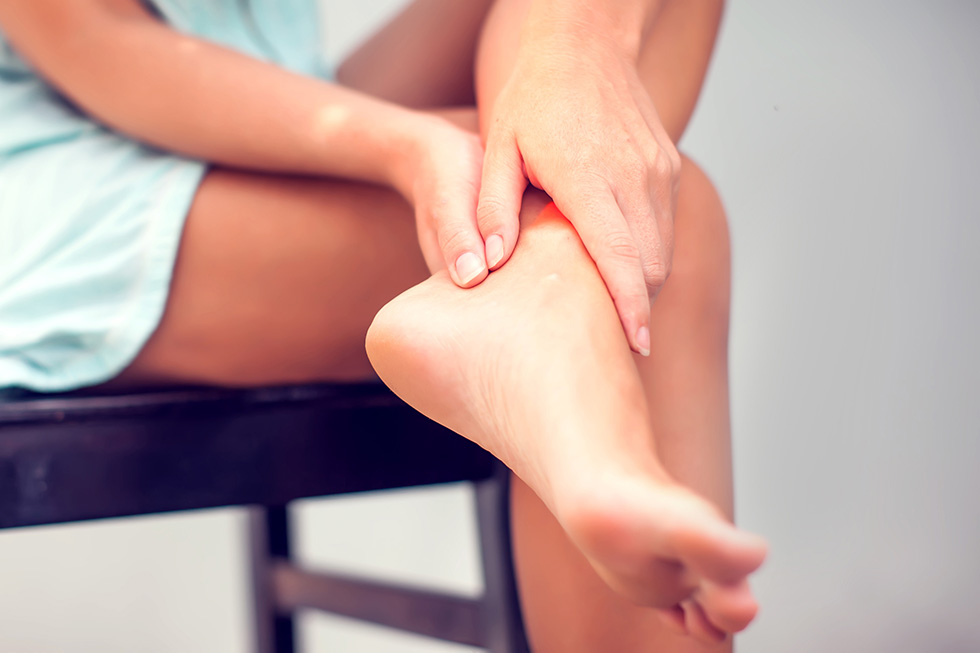 We provide comprehensive, conservative care for a wide variety of foot and ankle conditions.
 We offer same- and next-day care to patients with acute injuries.
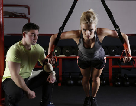 Get back in the game with help from our sports medicine specialists.
 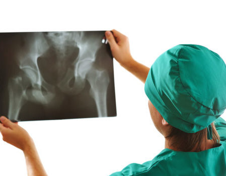 Our centers are equipped with a state-of-the-art digital X-ray machine.