


The Rotator Cuff: Impingement Syndrome and Rotator Cuff Tears
Aug 22, 2019Ty E. Richardson, M.D.
I. Anatomy
The rotator cuff is comprised of four separate muscles. The two most commonly involved tendons in rotator cuff pathology are those of the supraspinatus and infraspinatus. Both these tendons insert into the greater tuberosity. When viewed grossly, they are found to perform a continuous layer at the insertion. Two other important soft-tissue structures involved in impingement syndrome and rotator cuff tears are the coracohumeral ligament, and the long head of the biceps tendon. The function of the rotator cuff is to provide direct joint compression, which opposes superior migration of the humeral with forward elevation and abduction of the upper extremity. The rotator cuff also contributes significantly to abduction and external rotation strength.
II. Bony Anatomy
It is essential to understand the bony anatomy of the shoulder to understand the origin of impingement syndrome and rotator cuff tears. The most important structure is the anterolateral acromion process. Three distinct types of acromion have been described based on cadaveric studies. Type I is a flat-shaped acromion. Type II has a gentle curve. Type III has a very prominent anterolateral hook. The Type III is found in around 39 percent of the population. It is important to note that in cadavers with a Type III acromion, 70 percent were found to have a full-thickness tear of the rotator cuff.
III. Impingement Syndrome
Impingement syndrome has been called by several other names, mainly rotator cuff tendinitis or shoulder bursitis. This refers to chronic irritation of the tendons of the rotator cuff and the subacromial bursa due to bony impaction between the anterior margin of the acromion and the greater tuberosity of the humeral head. Damage to the rotator cuff follows a predictable course and is both age and activity related. Trauma is another important source of rotator cuff pathology. Incidence of rotator cuff tears with dislocation increases with age, and in those patients greater than 60 years of age with an anterior dislocation, more than 80 percent have traumatic rupture of the rotator cuff. The natural history of rotator cuff pathology does demonstrate some definite age-related changes. MRI’s performed on asymptomatic patients greater than 60 years of age revealed that 14 percent showed a full-thickness tear of the rotator cuff and 20 percent showed partial thickness tears.
IV. History
Patients often complain of a dull, achy pain, which is located at insertion of the deltoid in the mid lateral upper arm. This pain is exacerbated by overhead work, throwing or heavy lifting. One very common complaint is that of pain that awakes the patient from sleep. Weakness and decrease of active range of motion also accompany rotator cuff pathology, and the dominant arm is involved in 78 percent of these patients.
V. Physical Examination
Observation will often reveal atrophy of the supraspinatus and infraspinatus fossae. The patient will often have tenderness over the greater tuberosity. The patients with impingement will often have full range of motion, but pain in the extremes of forward elevation, which is referred to as the Neer impingement sign. Patients with a full-thickness tear will often have a markedly restricted range of motion and weakness. The inability to support their arm at 90 degrees of abduction in the scapular plane is called the drop-arm test, and this is indicative of a full-thickness tear.
VI. Radiographic Evaluation
Two radiographic views are usually sufficient to define the bony anatomy of the shoulder. The first is called the outlet view, and it is performed in the plane of the scapula, which clearly delineates the shape of the acromion. The anterolateral hook of a Type III acromion is easily seen on this view. The second view is called the Alvis view, which is the “Anterolateral View of the Impinging Structures”. This is performed at 25 degrees of external rotation at a 30 degree caudal angle. This shows the anterolateral margin of the acromion and profile, clearly demonstrating any anterolateral spurring of the acromion. MRI’s are between 96 to 100 percent sensitive for tears of the rotator cuff and can also display bony impingement. For patients who are unable to undergo an MRI, the arthrogram, which involves injection of a contrast material into the articular surface of the shoulder, is also very sensitive in demonstrating full-thickness tear rotator cuff tears.
VII. Treatment
Conservative care for impingement syndrome and rotator cuff tears consists of anti-inflammatory medication, subacromial injection of steroids and local anesthetic, physical therapy for strengthening of the rotator cuff and deltoid, and avoidance of offending activities, mainly overhead work. In the absence of cuff tears, properly performed subacromial injections are often 80 percent effective in relieving the pain of impingement. The surgical treatment of impingement syndrome is directed toward removal of the impinging spurs of the anterolateral margin of the acromion and the undersurface of the acromioclavicular joint. This is easily performed arthroscopically through multiple small incisions allowing rapid rehabilitation. Small full-thickness tears of the rotator cuff are also amenable to arthroscopic repair, however, large or retracted tears, which are not easily mobilized, are best treated through and open technique. The postoperative course after either arthroscopic or open repair involves very slow mobilization and passive range of motion for the first six weeks. Patients are usually able to return to light-duty work in eight to ten weeks. Those involved in strenuous occupations such as construction are often delayed as long as four to five months before their return to full work. Pathology of the rotator cuff continues to be a very challenging entity, both diagnostically and therapeutically.


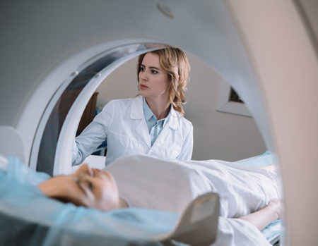 Our patients can receive MRI imaging onsite at both our Louisville and New Albany Clinics.
 Providing the latest advances in orthopedic surgery is our specialty.
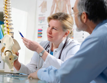 We take a unique, multidisciplinary approach to pain management.
 Our physical therapists use advanced techniques to help restore strength and mobility.
 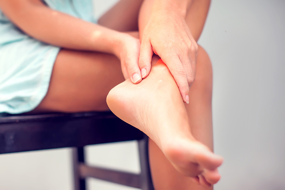 We provide comprehensive, conservative care for a wide variety of foot and ankle conditions.
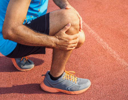 We offer same- and next-day care to patients with acute injuries.
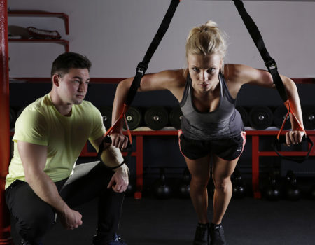 Get back in the game with help from our sports medicine specialists.
 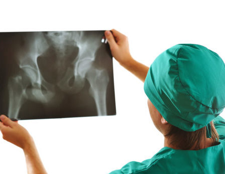 Our centers are equipped with a state-of-the-art digital X-ray machine.