The Arthritic Knee: Treatment Options
Aug 22, 2019Richard A. Sweet, M.D. (Retired 2022)
Introduction:
Total knee replacement (TKR) surgery has, along with hip replacement surgery, become one of the great medical and surgical success stories of the last several decades. Hundreds of thousands of patients each year benefit from these procedures. The number of knee replacements performed each year worldwide and in the U.S. climbs. This is due to a number of reasons. Our “baby-boom” population is aging. We are attempting and expecting to maintain active lifestyles much later in life than previous generations. As athletics have grown in popularity, the incidence of arthritis due to previous injury has climbed. Americans are indulging in an ever increasing daily dietary caloric intake, and resultant obesity adds stress to the weight bearing joints. This additional joint stress is contributing to the number of Americans who develop knee arthritis leading to replacement surgery.
Arthritis – General Concepts:
Arthritis is defined as the deterioration of the articular or surface cartilage of a joint. This progressive loss of cartilage is associated with inflammation, joint swelling and fluid, and painful range of motion. There are many types of arthritis. The underlying cause is generally unknown. There are two broad classifications of arthritis. These include inflammatory and non-inflammatory arthritis. The former is typified by the condition known as rheumatoid arthritis. The latter is typified by the type of arthritis known as osteoarthritis.
By far the most common type of arthritis is osteoarthritis, also known as degenerative arthritis. It is the type associated with aging and “wear and tear”. Its underlying cause is unknown. It is associated with a characteristic x-ray appearance (see below). Lab and blood work are non diagnostic (i.e. there is no positive blood test for this most common type of arthritis). The diagnosis is made on clinical grounds. Post-traumatic arthritis is a variant of osteoarthritis. It is distinguished by a previous history of injury, which has led to the development of the arthritic condition. It otherwise is usually similar in clinical appearance to osteoarthritis.
Inflammatory arthritis is much less common. Though inflammatory arthritis is typified by rheumatoid arthritis, there are other variants. Arthritis associated with other systemic conditions such as psoriasis, ulcerative colitis, lupus and others is of the inflammatory type. This type of arthritis is associated with an autoimmune disorder where the patients own immunologic system attacks the cartilage cells destroying them and creating a painful, inflamed, swollen, and sometimes deformed joint. Blood work is often diagnostic for these conditions.
Nonsurgical Care:
Nonsurgical management of arthritis should always be considered first. A combination of short periods of rest, ice/heat therapy, physical therapy, medical management, and exercise in moderation can be attempted. Success largely depends on the severity of the underlying arthritic condition. As the joint gets closer to “bone on bone”, all conservative measures become less effective.
NSAIDS
The use of oral nonsteroidal anti-inflammatant (NSAIDs) has been a standard part of the medical management of arthritis for years. NSAIDs are not disease altering. They neither “cure” nor even slow the progression of the arthritic process. They can, in early stages of arthritis, diminish the inflammation and pain of arthritis. In more severe stages of the disease, NSAIDs are less effective. The risk of gastrointestinal problems (gastritis or ulcer disease) has long been well known. There is now conflicting evidence regarding the cardiovascular safety profile of all NSAIDs, and it is recommended that prior to using these medications, medical consultation is obtained.
Steroid (Cortisone) Utilization
The oral use of cortisone (a form of steroid) can also be effective in controlling the symptoms of arthritis. Like NSAIDs, steroids are not disease altering. When used judiciously, they can be quite effective in reducing the pain, swelling and inflammation of arthritis. When steroids are over used, many complications can occur as almost every organ system in the body is affected by their use. This does not mean oral steroids should be completely avoided, but simply that their use should be monitored and controlled. Oral steroids are often given in a tapering dose pack taken over a week’s time, such as with a Medrol Dosepak.
Steroid (cortisone) injections can also be effective in relieving the pain, inflammation and swelling of arthritis, especially in its earlier stages. Excessive use of intra-articular steroid injections can cause accelerated deterioration of the arthritic joint. Though there are no absolute rules, the general recommendation is to not exceed three injections into a single joint over the course of a year. Exceptions can be made in circumstances of poor overall health or in the face of surgical contraindications.
Visco Supplementation Injections
Another conservative option is the use of “visco supplementation injections”. Visco supplementation is the process of injecting, by a series of three weekly shots, hyaluronic acid into the affected joint. Hyaluronate is the chemical found in normal fluid of the joint. It is theorized that injecting hyaluronate into the joint will improve the joint lubrication leading to a subsequent decrease in inflammation and pain. These injections are also popularly known as “gel” or “lubrication” injections. Several products are available commercially. These include Synvisc, Hyalgan, and Supartz. Though the manufacturer of each claims benefit for their particular product, there is no clear evidence demonstrating that one is more effective than the other. When hyaluronate injections were first released a few years ago, there was much hype and media fanfare. Many misinformed patients were led to believe that this would be a miracle cure for arthritis. Time has shown that while effective, visco supplementation injections are probably no more beneficial than steroid injections. Their advantage is the lack of side affects. The disadvantages include increased cost (often necessitating insurance company pre-approval), the delay in onset of benefit (steroid injections work in a few days while hyaluronate injections begin to work in two to three weeks), and the inconvenience of returning to the doctor’s office at weekly intervals to receive the three injections. As with steroid injections, the more severe the arthritic condition, the less effective will be the treatment. And, as with steroid injections, any benefit is temporary, lasting anywhere from a few weeks to several months depending on underlying severity of the arthritic condition.
Chondroitin / Glucosamine Products
Over-the-counter dietary supplements have become popular in treating the symptoms of arthritis. These are non FDA controlled substances for which there is “testimonial evidence” only supporting their effectiveness (unproven by rigid scientific, medically accepted and controlled studies). The most popular of these are the glucosamine and chondroitin products. Medically accepted studies are now underway to determine the effectiveness of these product’s claim to restore cartilage to the arthritic joint. Though there is some skepticism regarding these product’s effectiveness, they do appear to be safe and side effects appear to be rare.
Arthroscopy:
Arthroscopic “clean out” is a treatment option in caring for early arthritis of the knee. It has both benefits and limitations. Arthroscopy’s chief benefit is its outpatient percutaneous less-invasive nature allowing for quick recovery from surgery. Arthroscopy can resolve symptoms due to coexisting meniscal cartilage tears. Loose cartilage particles floating free in the knee that are produced by the arthritic process of the wearing down of the joint surface are irritating to the knee (causing it to swell or fill with fluid). These can be arthroscopically flushed from the joint. Larger loose fragments of bone and cartilage (loose bodies) can also be removed. These loose bodies can interfere with the smooth functioning of the knee joint and cause a painful “catching” sensation. Irregularities of the joint surface can also cause mechanical symptoms such as “catching”, pain and giving way. These areas can be smoothed, thus eliminating the mechanical symptoms.
While all of these benefits are intuitive in their potential to improve the clinical function of the knee, it needs be clearly pointed out that arthroscopy does not “cure” arthritis or even slow its progression. It does not restore cartilage to the joint surface. As with the use of NSAIDs, the procedure is not “disease altering”. And once the joint space starts to significantly narrow on a weight bearing x-ray, an arthroscopic clean out is seldom successful. Many surgeons, trying to help patients get a handle on the prospects for success of an arthroscopic washout of the arthritic knee, tell patients that there is a 50% chance of symptom reduction and a 0% chance of a cure.
Total Knee Replacement (TKR)
Surgical Indications:
The primary indication for total knee replacement (TKR) surgery is disabling pain. When more conservative measures can successfully control symptoms, TKR surgery should not be entertained as a treatment option.
Patient age plays a role in determining if TKR surgery is indicated. There are issues of wear and loosening, which make replacement surgery less attractive to younger patients (see “IMPLANT LONGEVITY” below). There is no absolute minimum age. The ideal candidate is over 60 years of age. However, many patients between 50 and 60 undergo knee replacement when the arthritic disease is truly end-stage and symptoms are intolerable for activities of daily living. Thorough counseling is required to determine if TKR surgery is appropriate in this age group. For patients under 50 years of age with severe arthritis, TKR surgery can still be considered. In this age group, however, the risks of cementless implant fixation or a hybrid fixation combining cement and non cement techniques becomes more attractive and can be considered as a surgical strategy to try to eliminate the risk of ultimate cement fixation loosening that will inevitably occur.
X-Ray Changes:
X-rays of the knee in patients requiring knee replacement are diagnostic. The chief x-ray finding in the osteoarthritic or post-traumatic knee includes loss of the joint space so that the knee is virtually rubbing “bone on bone”. Articular cartilage does not show up on x-rays. In the normal knee with thick, healthy articular cartilage, the articular cartilage shows up as a smooth symmetrical wide “joint space”. In the arthritic knee, the cartilage is worn or thinned. This shows up on the x-ray as narrowing of the articular cartilage space. The narrower the space, the more severe the arthritic process. In most patients requiring TKR, the space has worn completely away, thus presenting the “bone on bone” radiographic appearance.
Other characteristic x-ray changes in osteoarthritis include loss of bone stock in severe cases (where the bone is actually rubbing away), deformity (typically a bowed or knock-kneed appearance), and bone spurs (called osteophytes).
X-rays of patients with rheumatoid or inflammatory arthritis will show the same joint space narrowing as seen with osteoarthritis. The major radiographic difference is the typical lack of bone spurs present.
Risks of Surgery:
All surgical procedures carry certain risks, and TKR surgery is no exception. Studies have shown that the more experienced the surgeon and the surgical team, the less the risk of complications.
Infection is the dreaded complication when inserting any foreign body, such as knee implants, into the human body. This is because bacteria adhere to the foreign body, which of course has no blood supply, making eradication of the bacteria very difficult. Prevention of infection is the key to success. Intravenous (IV) antibiotics are administered pre and post operatively. The wound is irrigated with an antibiotic solution during surgery, and strict sterile technique is utilized including the use of “space suits” for the surgical team. The infection rate has now been driven down to less than 0.5% with these stringent measures.
Thrombophlebitis (blood clots) can occur. Prophylaxis with one of a variety of options minimizes this risk. Coumadin, an orally administered blood thinner, is one of the most common agents used. It is typically given the night of the surgical procedure and given for a duration of two to six weeks. Other injectable blood thinning options are also effective and available (Lovenox and others).
“Minimal Incision Surgery” Indications
Many patients are candidates for incorporation of newer Minimal Incision Surgical (MIS) techniques. The cosmetic result of the more minimal approach is a much smaller skin incision, sometimes 4 inches or less in length in an ideal candidate. However, the major benefit of MIS knee replacement technique is not the improved cosmesis, but instead the enhanced protection of the underlying muscles, tendons, and ligaments during surgery. In specific, the quadriceps tendon (the major tendon just above the kneecap that connects the quadriceps muscle to the patella) and the quadriceps muscle itself are protected during the surgical dissection. This “quad sparing” approach allows for easier and earlier regaining of range of motion of the knee and return of the ability to straight leg raise. The net result is a somewhat quicker and easier rehabilitation program, with less postoperative pain and a quicker return to daily activities.
The ideal candidate for MIS is a relatively thin patient without severe deformity or contractures and with healthy skin. Relative contraindications to MIS knee replacement include very large knees due to obesity, severe deformity or contracture of the knee, skin conditions that could affect healing potential such as psoriasis, or any other patient with increased healing or infection risk. However, even in patients where true MIS techniques are relatively contraindicated, many of the principles can still be applied facilitating an easier recovery even in these circumstances.
Surgical Technique:
The skin incision is straight midline anterior (just down the front of the knee). Its length depends on how amenable the knee is to MIS techniques (see above), and can vary from four to six inches. The capsule of the joint is opened just medial to the patella (kneecap). The quadriceps mechanism is protected as much as possible to allow for quicker return to function. Once inside the joint, bone is cut from the distal femur (end of the femur or thigh bone) and proximal tibia (shin bone). The thickness of the bone cuts basically matches the thickness of the implants and is generally about one centimeter thick. Some adjustments in depth of bone cuts are made to account for individual patient anatomy, contractures and deformity. Instrument systems are available to accurately and reproducibly create the bone cuts to provide for proper alignment of the leg. Recent MIS trends have led to the development of smaller and lower profile instruments to allow the surgeon to perform TKR surgery through smaller incisions. The distal femur is shaped to conform to the implant anatomy anteriorly and posteriorly. Implants are sized based on an individual basis depending on the size of the femoral and tibial bone itself. Often the deep surface of the patella is removed to allow for its resurfacing (it is a misconception that the entire patella itself is replaced – only the deep articular surface is replaced). Ligaments are balanced so as to provide for supple range of motion while still providing sufficient stability to allow for an active lifestyle. It is the balance between achieving this suppleness while preserving stability that is the “art” of the science of knee replacement surgery.
Implant Fixation
Once all the bone cuts have been created, the knee implants are usually cemented in place. Cementless fixation, while common in hip replacement surgery, is seldom used in knee replacement except in very young patients. Potential candidates for cementless implant fixation include patients under 50 to 55 years of age who have healthy strong bone and who are willing to accept up-front risks of fixation failure so as to eliminate the later risk of cement loosening. To date, cementless fixation in TKR surgery is not 100% dependable and there is risk of lack of bony ingrowth into the knee implants, which can lead to early loosening. This lack of bony fixation rarely occurs in hip replacement patients, resulting in the common use of cementless fixation by hip surgeons. For these reasons, most TKR implants are cemented. The length of time that cement fixation will hold up prior to loosening depends on many factors. These include the quality of cement technique used at the time of surgery, the age and activity level of the patient, patient weight, the quality of bone and other factors. The general goal is to achieve fixation via cement for 15 years or more.
After all implants are cemented into place, a trial reduction is preformed with varying thickness and levels of conformity of the polyethylene (plastic) insert. This provides for final adjustment of ligament balance and tension. Wound closure is meticulous to prevent problems in healing. A drain is sometimes inserted into the knee to be removed the first day after surgery.
Pain Management
Pain management is an important part of caring for a knee replacement patient. Several options are available to supplement the use of routine postoperative IV, intramuscular (IM), or oral narcotic pain medication. Regional nerve blocks can be preformed by the anesthesia team. Nerve blocks are typically performed either in the operating room just before or after surgery, or in a special holding room prior to surgery. These can, in varying degrees, “numb” the leg for the first 24 hours postop. The most common regional nerve block utilized is the femoral nerve block. The femoral nerve is injected with an anesthetic in the groin area. Though providing significant pain relief, the femoral nerve block alone does not completely anesthetize the leg. In conjunction with the femoral block, the sciatic nerve can be blocked as well. The sciatic nerve is blocked via an injection into the buttock area. The combination of the femoral and sciatic nerve block provides the most complete anesthesia for the immediate postoperative period for the patient.
Other more traditional methods of controlling pain are also utilized. The PCA (patient controlled analgesia) route of IV administration via a pump is popular. With this method, the patient actually presses a bedside button on the PCA pump that then delivers a small dose of narcotic through the IV tube. This can be repeated every six to eight minutes until pain relief is obtained. There is a preset lockout limit on the amount of narcotic that can be administered each hour. Though use of the PCA pump sounds intuitively attractive, some surgeons have found that standard IM injections administered by the nursing staff every three to four hours provides more complete pain relief than does the PCA pump method. And many patients find that strong oral narcotics such as oxycodone (Percocet, Percodan, Tylox, Endocet and others) given one to two tablets every three to four hours often is nearly as effective as the IM or IV methods of pain control.
Postoperative Rehabilitation in Hospital
Rehabilitation will start soon after surgery. Depending on the time of day of surgery, the complexity of the surgery and other individual issues, the formal rehab program usually begins the day of surgery or the first day postoperatively. Use of the continuous passive motion machine (CPM) is routine. This machine will typically be utilized for one to two hours twice each day. The nursing staff will, if possible, get each patient up in a chair the evening of surgery and perhaps even walk a few steps to the door or bathroom. The day after surgery (postoperative day one) a more formal therapy program is initiated. This involves further efforts at gaining range of motion, mobilization to reestablish walking ability, and early efforts to strengthen the leg musculature. Typically the patient may weight bear as tolerated (apply as much weight as they are able to withstand) from the outset. The goal of the initial in-hospital rehab program is to obtain 90 degrees of knee flexion and full extension to 0 degrees, have sufficient muscular control so as to be able to straight leg raise, be able to walk well supported by the use of a walker or crutches, and be able to manage stairs. Speed with which these goals are met depend on many factors. The less knee motion available preoperatively, the harder it will be postoperatively to regain supple range of motion after surgery. For instance, the presence of a severe flexion contracture preoperatively makes gaining full extension to 0 degrees more difficult. Healthy, robust patients regain functional walking ability more quickly than those with less energy and vitality. Individual patient determination and ability to work through the normal postoperative pain affects the speed with which the postoperative goals are met.
Hospital discharge date also varies depending on an array of factors. Young, energetic patients who have undergone MIS can often be discharged on the second postoperative day. Elderly patients planning on a return to their home environment may, in some cases, be discharged from the hospital on the fourth postoperative day. The majority of patients leave the hospital on the third day postoperative.
After Hospital Rehabilitation
All patients require continued therapy after hospital discharge. Three basic options are available. These include 1.) transfer to a rehab facility, 2.) home therapy and 3.) outpatient therapy.
Rehab Transfer
Many excellent inpatient rehabilitation facilities now exist. Their development has been fueled by the demand by insurance carriers and Medicare to shorten hospital stays to better control costs. A patient is a candidate for such an acute-care rehab facility if it is anticipated that the need for rehab admission will be relatively short (10 to 14 days typical). Bed availability is sometimes tight, and advance bed reservation made at the time of scheduling surgery helps, but does not guarantee admission. Insurance coverage for rehab facility stays varies from one carrier to another. Medicare generally does cover admission. Younger patients are typically not covered, but seldom need such inpatient care in any case.
Home Therapy
Home therapy is a very popular post-hospital discharge option. The use of home therapy requires some family support at home. Typically a therapist will come to the home daily for one week then three times a week thereafter. A nurse will also come by and check each patient to draw any necessary blood work, check the incision area, and monitor the overall medical status of each patient. The home health industry has become much more sophisticated in recent years, and although the physical therapist does not have all the equipment that would be available in an outpatient or rehab setting, the quality of therapy is now excellent and many patients achieve excellent results. Home therapy is utilized for a few weeks postoperatively. When a patient is more mobile a switch to outpatient therapy is sometimes necessary if all rehab goals have not been met.
Outpatient Therapy
Though less convenient than home therapy, outpatient therapy has certain advantages. Not insignificantly, the actual process of getting dressed, out the door, into a car, out of the car, into the therapy department and then to reverse the process on the return home is therapy in and of itself. Once in the therapy department, special equipment is available to assist in rehabilitation. Typically a patient choosing this route (often younger, more mobile patients) will visit the therapy department daily for one week then three times a week thereafter until rehab goals are met (usually four to six weeks). Many outstanding outpatient physical therapy department options are available.
Expected Postoperative Milestones
As a patient progresses through the postoperative regimen, certain milestones are reached. Hospital discharge is on the second to fourth day postop. The average length of time on a walker or crutches is two to three weeks. In uncomplicated cases the patient may progress off the walker as able and in some cases, especially when MIS techniques have been employed, be off the walker a week or two after surgery. Most patients require the use of a cane in transition to independent ambulation. By four to six weeks most patients have reached this milestone of independence.
A patient can typically return to a sedentary job six weeks postoperatively and a physical job three months after surgery. There is a considerable range in patient’s individual ability to tolerate residual soreness and motivation to return to work making the return to work milestone quite variable.
By six months postoperatively almost all residual soft tissue soreness has subsided. Occasionally a patient will exhibit mild residual soreness for up to a year after surgery.
With regard to recreational sports, light athletic activities, such as golf, can be enjoyed three months after surgery. More strenuous sports, such as double tennis and snow skiing, usually require four to six months till the patient is sufficiently comfortable to participate.
Ultimate Expected Function of a Replaced Knee
The ultimate goal upon full recovery after TKR surgery is to achieve a painless functional joint for activities of daily living. Light recreational sports such, as golf and walking, are well tolerated. The knee will usually tolerate slightly more strenuous sports, such as doubles tennis and snow skiing. Though the replaced knee may tolerate more strenuous sports still such as jogging/running and single tennis, there are concerns regarding excessive wear on the joint leading to early revision.
Ultimate range of motion goals utilizing new surgical attempts to obtain “deep flexion” is from full extension to 125 degrees or more of flexion. Many factors can affect the final degrees of flexion achieved. These include the size or girth of the knee. The larger the girth the harder it is in the beginning stages of rehab to bend the joint due to the early impingement of freshly operated and sore tissues in the back of the knee. Poor preoperative motion makes gaining deep flexion after surgery more difficult. Patients who tolerate pain poorly and do not enthusiastically pursue the rehab program do not end up with deep flexion.
Implant Longevity
The biomaterials used in TKR surgery are increasingly durable. However, as with all man made mechanical moving parts, wear is inevitable. There are two common methods by which an artificial knee will begin to have problems due to wear. These are loosening of the implant from the bone and wearing down of the polyethylene (plastic) articular bearing surface.
Polyethylene Wear
In a well-performed knee replacement with the ligaments balanced and the bone cuts accurately aligning the leg, the polyethylene implant should last 10 to 15 years. This time period will depend in part on the patient’s weight and level of activity. When the polyethylene fails by a wear mechanism, a relatively straight forward operation can be preformed to replace it with a new thicker polyethylene without changing the femoral or tibial implants if they are still well fixed to bone.
Implant Loosening
The great majority of implants are fixed to the bone by using a cement called polymethylmethacrylate. Cement is used for its reliability and predictability in routinely producing excellent surgical results. The major drawback to cement is that given enough time, it will eventually crack and begin to loosen from the bone. The length of time the cement will hold the implant tightly bonded to the bone depends on many factors. One of the most important is the technical quality of cementing performed at the time of surgery. Surgeon experience and attention to well-established surgical principles will prolong the life of the cement bond. Again, patient factors are important as well. Obesity and a strenuously active lifestyle put extra stress on the cement. The goal is to obtain implants that will remain well fixed for 12 to 15 years or more before clinically significant loosening occurs leading to the need for revision surgery. When implants do loosen, revising them is possible, but the surgery is more demanding and the potential for complications higher.


 Our patients can receive MRI imaging onsite at both our Louisville and New Albany Clinics.
 Providing the latest advances in orthopedic surgery is our specialty.
 We take a unique, multidisciplinary approach to pain management.
 Our physical therapists use advanced techniques to help restore strength and mobility.
  We provide comprehensive, conservative care for a wide variety of foot and ankle conditions.
 We offer same- and next-day care to patients with acute injuries.
 Get back in the game with help from our sports medicine specialists.
 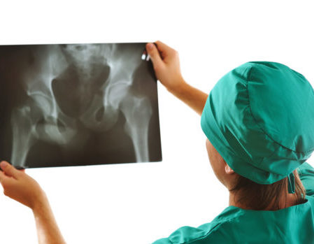 Our centers are equipped with a state-of-the-art digital X-ray machine.