


Distal (Non-Chevron) Metatarsal and Proximal Phalanx Osteotomies
Aug 21, 2019George Edward Quill, M.D. (Retired 2023)
As is true in all other areas of orthopaedics, appropriate patient selection is the key to obtaining a successful outcome when performing distal osteotomies for hallux valgus. Patients who either have significant pain and/or progression of deformity enough to warrant surgical correction by these techniques should probably not have a hallux valgus angle of any greater than 30 degrees, and ideally it should be passively correctable to the midline on preoperative physical exam. Distal osteotomies of the first metatarsal will usually only achieve satisfactory correction of a first intermetatarsal angle if it is less than or equal to about 15 degrees. One would have to imply different techniques for deformities with angles greater than this. It goes without saying that the patient should have a very good range of motion at the hallux MP joint to consider for a distal osteotomy.
The patient with generalized ligamentous laxity or hypermobility of the first ray would be prone to recurrence of hallux valgus if managed with only a distal osteotomy, and a more proximal procedure should be planned for those patients. Again, the two main indications for considering hallux valgus surgery are pain and/or progression of deformity. When employing a distal osteotomy, one must beware of the typical Morton’s foot that has a very long second ray relative to the first.
As early as the 1880’s Reverdin and Barker described an intra-articular, medially-based closing wedge distal first metatarsal osteotomy for hallux valgus. In two separate reports in the 1920’s, Homan described an extra-capsular, medially-based closing wedge distal first metatarsal osteotomy, in which he displaced laterally the capital fragment and tilted it plantarward. In 1945 Hawkins, Mitchell and Hedrick slightly modified the Homan technique to give us what we commonly refer to today as the Mitchell osteotomy. This is a lateral displacement angulation osteotomy of the distal first metatarsal fixed with sutures and accompanied by medial eminence resection and plication of the medial capsule. Gibson and Piggott described a peg-in-hole type of distal metatarsal osteotomy that is fairly similar to the Mitchell procedure, but these authors left the lateral plantar spike on the proximal fragment of the first metatarsal. They then impaled the distal metatarsal head fragment on this spike, thus the peg-in-hole description.
In 1925, Akin suggested a varus producing osteotomy of the proximal phalanx combined with medial eminence excision and medial capsular plication. For the relatively unrelated problem of hallux rigidus, Moberg described a dorsiflexion osteotomy, closing wedge, on the dorsum of the proximal phalanx of the great toe.
The indications for the Reverdin osteotomy are that the hallux valgus angle not be any greater than 30 degrees and the IM angle be less than 15 and preferably closer to 12 degrees. The patient would have to have very good range of motion of the hallux MP joint because this is an intracapsular osteotomy, and the dissection can lead to stiffening of the joint. This type of distal intracapsular osteotomy is contraindicated in a patient with an already short first metatarsal or the patient with hallux valgus interphalangeus. Osteotomy of the metatarsal wouldn’t necessarily correct the interphalangeus problem.
The technique for the Reverdin osteotomy includes a straight longitudinally oriented medial incision. I usually perform a Y-shaped capsulotomy with the broadest portion of the Y based distally, and attached to the proximal phalanx. The medial eminence is resected along the parasagittal groove of the first metatarsal, and this medially-based osteotomy is made in the cancellous bone at the distal end of the first metatarsal. The bone wedge is removed, and the osteotomy is closed. This can be fixed according to the surgeon’s preference and a medial capsulorrhaphy done with imbrication, first excising the redundant capsule.
The advantages of the Reverdin osteotomy are that it is a relatively simple technique and that the procedure may actually lessen the metatarsus primus varus if the first intermetatarsal angle is greater than 11 degrees preoperatively. Reverdin, as well as Mann and Coughlin, have made the observation that any procedure for the hallux valgus-metatarsus primus varus complex that corrects and maintains the correction of the excessive valgus of the hallux will reduce the first intermetatarsal angle. The disadvantage of this procedure is that it may actually worsen metatarsus primus varus if one isn’t careful and that it does indeed shorten the first metatarsal a certain distance. It may actually stiffen the hallux metatarsophalangeal joint because it is intracapsular, and most of these patients lose some range of motion in plantar flexion. Hallux valgus does recur in a significant number of patients.
The Mitchell osteotomy is indicated for patients with a hallux valgus angle of less than 30 to 35 degrees and again an intermetatarsal angle no greater than 15 and preferably closer to 12 degrees. The patient must have good range of motion at the hallux MP joint. This procedure may indeed correct mild to moderate degrees of incongruity. This osteotomy again is contraindicated for patients who have a short first metatarsal relative to the second and to those who have hallux valgus interphalangeus. If the patient is unable to protect weight bearing for whatever reason, you may want to choose a different osteotomy because displacement of this osteotomized segment of the first metatarsal can lead to disastrous results.
The technique for the Mitchell osteotomy again includes a medial approach to the first metatarsophalangeal joint or Y-shaped capsulotomy based distally and medial eminence resection. One must be careful not to dissect the lateral capsule off the metatarsal head and neck in order to preserve its contribution to the blood supply of the metatarsal head. Two holes are drilled perpendicularly through the metatarsal shaft from dorsal to plantar, making the holes just large enough to allow passage of a #1 suture. The first hole is about 15 millimeters proximal to the articular surface of the metatarsal head and oriented on the medial side of the head. The second hole is about 1 centimeter proximal to the first hole, but displaced laterally more towards the center of the head and neck. A #1 suture is passed through the hole so that it can be tied dorsally later. A double osteotomy of the metatarsal neck is then done, beginning 3 to 4 millimeters proximal to the distal hole. This osteotomy is made with a very thin saw blade and is incomplete and doesn’t go through the lateral cortex, leaving 3 to 6 millimeters of the lateral shaft intact. The width of the lateral spike thus created will depend upon the amount of correction needed to relax the lateral soft tissue of the joint and dictates how far the distal fragment will be laterally translated. The second osteotomy is perpendicular to the long axis of the metatarsal shaft, starting on the medial side 3 to 4 millimeters proximal to the first cut. This second osteotomy, which is more proximal, is complete through the lateral cortex. These osteotomies can be diverged plantarward about 10 degrees to facilitate plantar flexion of the distal fragment. The capital fragment is displaced laterally.
The advantages of the Mitchell osteotomy are that it is very well studied and it has been proven that there is a less than 1 percent nonunion rate. Recurrent hallux valgus is rare, and this can indeed correct the appropriate amount of deformity and narrow down the forefoot. The main disadvantages of this procedure are that there is a very high rate of metatarsalgia, due to either dorsiflexion malunion, shortening of the first metatarsal, or both of these factors. This is also considered to be a very technically demanding procedure.
One can remember certain perils when performing the Mitchell osteotomy. The osteotomy must be made in cancellous bone so that it will heal faster. The suture must be protected during the osteotomy so that it is not cut by your saw blade. You want to avoid an uneven lateral spike on the distal fragment because this may also result in instability. Take care to align the first metatarsal head with the second and avoid dorsal angulation or dorsal displacement, trying not to strip the lateral capsule because of the risk of avascular necrosis.
The Akin osteotomy can be employed in a number of situations, usually in conjunction with another reconstructive or realignment procedure.
The best indication for this procedure is the correction of hallux valgus interphalangeus. The laterally inclined hallux MP joint, which is indeed congruent in a valgus position, can be corrected with periarticular osteotomies, including the Akin osteotomy. This may also be used as a salvage procedure to realign the great toe if it is stiff or if there has been failure of prior procedures.
This procedure is rarely used alone for hallux valgus and most commonly is used in patients greater than 45 years old with a hallux valgus angle somewhere between 30 and 50 degrees. It is usually best if the IM angle is less than 12 degrees.
The technique for the Akin osteotomy involves medial eminence removal and adductor tenotomy. A basilar osteotomy is made in the cancellous extra-articular portion of the proximal phalanx. It is a medially based closing wedge osteotomy. It is usually best if the surgeon does not go all the way through the lateral cortex when making the saw cut. The wedge is removed, and the deformity is corrected. The surgeon may either chose to fix the osteotomy with pins or sutures or may rely upon soft tissue closure. A hint here would be to use a nonabsorbable suture rather than a stainless steel wire.
The advantage of the Akin procedure is that it may be combined with other hallux valgus procedures to affect a good result. Other procedures include metatarsal osteotomy, adductor tenotomy, and medial eminence resection. This is a good procedure for the laterally-inclined hallux MP joint and for salvage of recurrent hallux valgus. It is usually best applied for the patient with hallux valgus interphalangeus.
The following is an example of a patient whose hallux valgus deformity was managed with an Akin osteotomy.
The disadvantages of this procedure are that it rarely works when it is used as the sole reconstructive procedure. Fixation and pin problems are common, and usually the clinical picture looks much better than the x-ray appearance.
The Moberg osteotomy is a dorsally-based closing wedge osteotomy of the proximal phalanx used to increase dorsiflexion in cases of hallux rigidus and hallux limitus. This actually changes available plantar flexion range of motion to dorsiflexion at the hallux metatarsophalangeal joint.
Bill Hamilton has coined some interesting terms and essentially invented a new nomenclature for other osteotomies about the forefoot, and he has been kind enough to supply me with the following slides. A Nika osteotomy is actually a reverse Akin procedure that is done for metatarsophalangeal varus. This is a laterally-based closing wedge of the proximal phalanx used to straighten the toes out of varus.
An Akinette procedure is very similar to the Akin procedure described above, but is performed on the fifth metatarsophalangeal area.


 Our patients can receive MRI imaging onsite at both our Louisville and New Albany Clinics.
 Providing the latest advances in orthopedic surgery is our specialty.
 We take a unique, multidisciplinary approach to pain management.
 Our physical therapists use advanced techniques to help restore strength and mobility.
 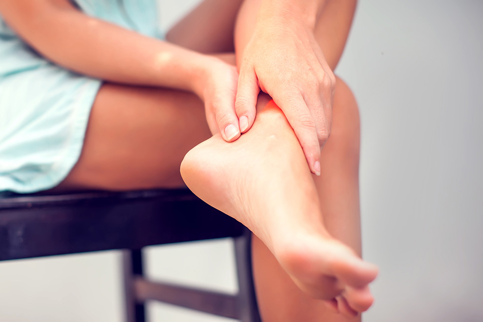 We provide comprehensive, conservative care for a wide variety of foot and ankle conditions.
 We offer same- and next-day care to patients with acute injuries.
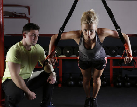 Get back in the game with help from our sports medicine specialists.
 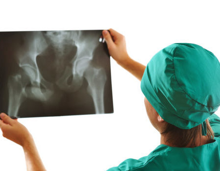 Our centers are equipped with a state-of-the-art digital X-ray machine.