


Total Knee Replacement: Deep Flexion
Aug 22, 2019Richard A. Sweet, M.D. (Retired 2022)
Introduction
Over the last three decades total knee replacement (TKR) surgery has evolved into a reproducibly successful procedure benefiting hundreds of thousands of patients each year. Greater understanding of proper implant design and standardization of surgical technique has occurred. And as the procedure has matured over the last decade, the pace of its evolution has slowed. However, two recent developments have quickened that pace of change.
The first of these is the development of more minimally invasive methods of performing TKR. Minimally invasive surgery (MIS) has benefited patients by diminishing immediate postoperative pain, shortening hospital stay and rehabilitation time, and overall making the TKR surgical experience easier to endure. To a large extent it has changed forever the way in which TKR surgery is performed and with it patients’ expectations.
The second of these developments has been the concept of providing for patients a prosthetic knee that allows for deeper flexion. TKR patients who have successfully achieved deep flexion can lead a more normal, easier and freer lifestyle having to make fewer minute to minute and day to day concessions to their artificial knee. Simple lifestyle events, such as putting on shoes and socks, foot hygiene, sitting in movie theaters and airplane seats, getting into and out of the back seat of cars, and getting into low spots around the home, all are made simpler and more comfortable when deep flexion in a TKR has been achieved.
General Concepts
Deep flexion TKR is arbitrarily defined as a knee that achieves flexion greater than 115 to 120 degrees. In the early development of the resurfacing TKR surgery, 90 degrees of flexion was considered sufficient or even ideal. Indeed, there was concern that with deeper flexion posterior instability would be risked and that polyethylene wear would be enhanced. Concerns regarding these issues still exist. These concerns are valid if the surgeon achieves deep TKR flexion without adhering to strict surgical principles regarding stability and proper tibiofemoral tracking. To achieve a high flexion TKR that is symptom free and stable requires a thorough understanding of normal knee kinematics including the concept of femoral rollback and the need for physiologic posterior stability. The goal of deep flexion TKR surgery is to obtain it while maintaining a balanced, kinematically functional, stable knee.
Kinematics in the Normal and TKR Knee
As the normal knee flexes, femoral rollback occurs. The lateral femoral condyle, having a larger radius of curvature, rolls back farther posterior than the medial femoral condyle.7 This rollback is guided by the posterior cruciate ligament (PCL).1 The asymmetric rollback results in the tibia internally rotating relative to the femur during flexion.
In the TKR patient, normal kinematics must also be guided by a functioning PCL. If the TKR is posteriorly unstable, paradoxical anterior slide of the femur on the tibia occurs and normal knee kinematics are not exhibited3 This paradoxical anterior slide of the femur on the tibia during flexion can be a cause of symptoms. These may include difficulty with stairs and inclines (particularly going down), soreness when the knee is flexed and loaded, such as with recreational athletic activities, and paradoxical anterior femoral slide on the tibia can be a cause of intermittent effusions as the femur repetitively stresses and irritates the anterior capsule of the knee. In addition, anterior sliding of the femur can cause earlier impingement of the posterior polyethylene on the back of the femur, thus preventing high flexion from occurring3.
To achieve a high-flexion, symptom-free knee, normal kinematics must be understood. It is not satisfactory to achieve deep flexion knee arthroplasty if it is posteriorly unstable and functionally symptomatic due to altered knee kinematics.
Implant Design Considerations
Patellofemoral Joint Design Features
To achieve deep flexion in TKR surgery, certain design principles are required of the implants. The location of the patellofemoral joint must be anatomic. Anteriorizing the patellofemoral joint, as occurs with many resurfacing TKR implant designs, puts excessive strain on the extensor mechanism preventing the knee from achieving deep flexion. Recessing the trochlear groove resolves this problem and can be managed via different design strategies. One method involves actually cutting a recessed groove into the chamfered femoral bone. This technique adds an additional step and instrumentation to the surgical procedure. A second simpler method involves simply making the femoral chamfer cut itself thicker while making the trochlear groove of the femoral component deeper.
Further, the femoral trochlear groove must extend far enough posterior so that with deep flexion the patella will not impinge in its depths and will instead ride congruently throughout the flexion arc. Otherwise a painful patellar “clunk” can occur as the patella catches into an insufficient trochlear groove when coming out of maximum flexion
Femoral Component Design Features
The femoral component should have a smooth radius of curvature transition throughout its entire flexion – extension arc. This rounded sagittal profile reduces soft tissue forces in mid flexion allowing for a smooth transition from extension to flexion.
Another important femoral component feature in providing for a deep flexion implant is the design of the posterior femoral condyle. Deep flexion requires the presence of a slightly thicker posterior condyle. The radius of curvature throughout this somewhat thicker posterior femoral condyle should be a continuation of the sagittal curvature arc present at earlier stages of flexion. This requires a somewhat thicker bony posterior condyle resection.2 A relatively thin and flatter posterior condyle design, as is present in many implants, results in early impingement of the tibial implant on the femur preventing further flexion. Also the posterior condyle needs to be of an appropriate length. If too long, it will overhang the under lying femoral bone and subject the femoral implant to levering forces in deep flexion. If too short, bony impingement can occur preventing deep flexion.
Femoral sizing is also important. If improperly sized, flexion will be compromised. If the femoral component is too large in a medial – lateral dimension, soft tissue irritation may limit the patient’s ability to gain flexion during the early rehab stages. If improperly sized in the anterior – posterior dimension, improper placement of the patellofemoral joint or over stuffing of the flexion space can also result in limiting flexion. Thus, the femoral component must be designed proportionally to proper medial/lateral versus anterior/posterior dimensions. A variety of femoral sizes need be available to fit all anatomic situations.
Tibial Component Design Features
In the posterior cruciate retaining knee, the tibial implant polyethylene design should allow for anatomic rollback guided by the posterior cruciate ligament.3 However, an excessively flat polyethylene design risks peak point contact stresses and posterior edge loading (if rollback is excessive) resulting in increased polyethylene wear.4 5 Thus, some congruence is required. In addition, due to the concerns regarding posterior instability in deep flexion, multiple polyethylene constraint options enhancing stability are necessary. Posteriorly lipped or dished implants, anteriorly lipped implants that are effective at enhancing stability when descending stairs, and various levels of PCL substituting implants should be available. “Anterior lipped” polyethylene inserts can theoretically be particularly effective in helping to prevent paradoxical anterior sliding of the femur on the tibia in flexion.
A variety of polyethylene constraint options should be available for the surgeon to intraoperatively dial in the level of congruence and restraint necessary to balance mobility and flexion versus stability.
Implant Design: Conclusion
If the TKR implants chosen by the surgeon are improperly designed, and if the surgeon still strives for a deep flexion performing knee, the only method of achieving this goal is to compromise knee stability. Thus, implant selection and design is crucial to the overall success of deep flexion TKR surgery.
Patient Factors
Patient factors affect the surgeon’s ability to achieve deep flexion. Obesity and increased overall girth to the knee have a negative impact on ultimate range of motion. This occurs for several reasons. First, the size of the knee itself can act as a mechanical block to further flexion. Second, in a freshly operated knee the posterior tissues are sensitive and sore. As the knee is flexed, these tissues in the popliteal space impinge upon themselves creating pain. The larger the girth of the knee, the earlier in the flexion arc this painful impingement occurs and the harder it is for the patient to overcome it, thus limiting ultimate flexion achieved.
Another patient factor limiting ultimate flexion achieved is the amount of preoperative flexion and range of motion available.2 6 It has long been known that the less motion a patient has prior to TKR surgery, the harder it is for the patient to achieve range of motion after surgery. A knee with a severe flexion contracture or one that will flex poorly preoperatively is unlikely to be a knee that achieves supple, stable deep flexion postoperatively. Similarly, a multiply operated knee is unlikely to realize deep post-TKR flexion. Patients with severe varus or valgus deformities are surgical challenges to achieve stable postoperative range of motion.
Surgical Technique
General Concepts
The surgical technique to obtain deep flexion in total knee arthroplasty follows standard exposure principles. MIS techniques can be utilized. When MIS technique is performed, flexibility in terms of the sequence of bony cuts is appropriate
Distal Femur & Proximal Tibial Cuts
The distal femoral cut and the proximal tibial cut depth need be of sufficient depth. If less bone is removed than thickness of implant replaced (except in cases of bone stock loss), over stuffing of the joint will occur and supple range of motion will not occur despite all other efforts. The surgeon’s instinct is always to preserve bone stock. But if insufficient distal femur and/or proximal tibia bone is resected and the knee is over stuffed, not only will a deep flexion knee not occur, but even marginally functional range of motion will not be achieved. Allowance should be made when evaluating the adequacy of the distal bone cuts for the thickness of cement to be later utilized.
Excessive distal femoral bone resection elevating the joint line can also be a cause of failure to achieve supple postoperative motion. This error creates a relative patella baja and abnormal patellofemoral joint forces limiting flexion. It should be noted, however, that resecting an extra two millimeters of distal femoral bone stock in the face of a significant preoperative flexion contracture, though theoretically raising the joint line slightly, can often help eliminate a flexion contracture without the need for major soft tissue releases. Minimizing the extent of soft tissue dissection in such a manner can cut down on postoperative pain and soreness, thus ultimately gaining range of motion.
Varus/valgus distal femur and proximal tibial bone resection errors can also affect ultimate range of motion of the knee by unbalancing the collateral ligaments. Prevention of this error is best performed by use of cutting blocks based off an intramedullary rod. Careful central positioning of the intramedullary rod in the distal femur and proximal tibia is important. Computer navigation may replace the intramedullary rod as the most accurate method of aligning distal femoral and proximal tibial bone cuts.
Similarly, improper anterior/posterior sloping of these bone cuts can have a negative impact on flexion and extension. If the femoral cut is in extension, posterior femoral condyle bone is thrown into relief after prosthetic implantation and can impinge posteriorly. Increasing the posterior slope of the proximal tibia cut makes obtaining flexion easier, but can lead to posterior and flexion gap instability if excessive. An error in the proximal tibia cut so as to create an anteriorly sloping proximal tibial surface tightens the posterior capsule and may lead to posterior tibial under resection and compromised flexion gap. Earlier impingement of the polyethylene on the back of the femur can occur as a result of cutting the tibial in extension. Again, intramedullary rod instrumentation and computer navigation systems help avoid these errors.
Femoral Condyle Posterior Cut
When sizing the femoral implant and planning the anterior and posterior femoral bone cuts, two strategic options are available. These include the posterior referencing system and the anterior referencing system.
Posterior referenced sizing and planning of the anterior and posterior cuts are preferred by the author. This technique removes a measured and exact thickness of femoral bone posteriorly that will later be replaced by the posterior condyle of the femoral implant. In this manner the flexion space is planned for and replaced with precision. Femoral posterior offset can be accurately maintained by precisely excising a measured thickness of bone and replacing it with the same thickness implant.
Precision in the posterior condyle cut and replacement is one of the most important concepts in obtaining a high flexion TKR. The most serious mistake is under resection of the posterior femoral condyle. This results in flexion space over stuffing and can lead to functionally severe loss of flexion.
Over resection of the posterior femoral condyle also has significant impact on the amount of flexion that can be achieved in the TKR. If too much posterior condyle bone is resected relative to the thickness to be replaced, reduced posterior femoral offset occurs. Decreasing the offset posteriorly has a dramatic influence on potential knee flexion. For every millimeter of offset lost, flexion can be reduced by 6 degrees.3 Offset reduction can be another cause of polyethylene impingement against the posterior aspect of the femur.3
Anterior referenced systems are superb at preventing anterior notching, but resect a varying thickness posterior femoral condyle making flexion space balancing less precise and exact. This can increase the risk of flexion space over stuffing, flexion space instability, and/or femoral offset reduction. It is important to note, though, that there are many surgeons and implant systems that achieve terrific results with anteriorly based systems.
Femoral Anterior Condyle Cut
The accuracy of the anterior cut is nearly as important as that of the posterior cut (hence some surgeons preference for anterior referenced femoral sizing and cutting systems). If too deep, the anterior cut will notch the femur risking later fracture. If too superficial and too little bone resected, the patellofemoral joint will be displaced anteriorly. Anteriorization of the patellofemoral joint creates excessive strain in the extensor mechanism robbing the knee of flexion.
Component Malrotation
Malrotating either the femoral or tibial components will affect the mechanics of the knee and rob the knee of flexion. Internal rotation of the femur, as can occur with posterior femoral referenced systems in valgus knees with lateral condyle bone stock loss, will excessively tighten the lateral patellofemoral ligaments. At best, a lateral patellar release adding to the morbidity of the procedure will be required. At worst, knee flexion will be limited and patellar instability may occur. Internally rotating the tibial component, a common error in Minimally Invasive techniques, can increase the “Q” angle of the patellar tendon leading to similar patellar mechanics and problems.
Release of Contractures
All contractures must be released and the soft tissues balanced to achieve the goal of deep flexion and a stable knee replacement. As a general rule, all bone cuts should be completed and osteophytes removed prior to considering the release of any soft tissues. In many cases, extensive soft tissue release can be avoided, thus minimizing immediate postoperative pain.
As mentioned, flexion contractures sometimes can be easily managed by taking two extra millimeters of bone when performing the initial distal femoral osteotomy. On trial reduction, if the flexion contracture still persists, further action is necessary. If the knee is tight in flexion, as well as in extension, simply resecting more tibia may solve the problem. If the knee is balanced in flexion, options to eliminate the persisting flexion contracture include 1) ensure complete posterior meniscectomies have been performed and that no posterior capsular soft tissues are excessively tight – if so release the tight capsule while always protecting the peroneal nerve, 2) ensure that all impinging posterior osteophytes, particularly on the distal femur, have been removed, 3) resect another one to two millimeters from the distal femur, and 4) recess the PCL.
In a knee with a persistent varus contracture, initially inspect the medial joint line. The deep medial capsular ligament should be completely freed from the tibia. All tibial osteophytes should be removed. If the varus contracture still persists, the superficial medial collateral ligament and capsule should be progressively released from the tibia starting anteriorly and progressing posteriorly frequently checking for balance to make sure over release does not occur. Rarely the PCL will require recession to correct a severe varus contracture.
Valgus contractures are more difficult and dangerous to manage due to the risk of peroneal nerve injury. Release of the popliteus tendon is usually required. Further soft tissues considered for release include 1) iliotibial band, 2) lateral collateral ligament, and 3) PCL. The likelihood of requiring a PCL substituting knee increases in knees requiring extensive release to balance a severe valgus contracture.
Patellofemoral joint balance is required as well to obtain maximum flexion. To ensure balance, check for symmetry of the patellar cut. Lateral facet under resection can over tighten the lateral patellar ligament. Excessive femoral component internal rotation (a difficult problem to correct) also can over tighten the lateral patellar ligament. An anteriorized patellofemoral joint, as mentioned, also over tightens and unbalances the patellar soft tissues. Any or all of these will rob the knee of deep flexion.
Tibial Trial “Liftoff”
Understanding the concept of and routinely checking for “tibial liftoff” is one of the most important steps in reproducibly performing deep flexion total knee arthroplasty. After all bone cuts have been made and the soft tissues have been balanced, a careful trial reduction is performed. The key is to perform this step using a tibial base plate that “floats” on the tibial cut surface. The patella must be reduced. As the knee is maximally flexed, the point at which the anterior tibial trial tray begins to lift off the front of the tibia is the maximum flexion a patient is likely to realize after rehabilitation is complete. Early liftoff usually implies either that the flexion space is too tight or that rollback guided by the PCL is excessive. If the knee is already in deep flexion when liftoff occurs, no further efforts need be made and the implants can now be cemented into position. If anterior tibial trial liftoff occurs short of this flexion goal, further steps must be taken. Steps to correct early liftoff include 1) deeper proximal tibial resection if knee is tight in extension as well, 2) increase of the posterior slope of the proximal tibial cut which will effectively increase the flexion gap as the PCL guides roll back, 3) shift the femoral component anteriorly by resecting more posterior femoral bone and using a smaller size femoral component, 4) PCL recession or resection to correct rollback if it is excessive, and 5) ensure all contractures are released.
Checking the tibial trial liftoff point on the flexion arc and making sure this occurs at a degree of flexion that is satisfactory to the surgeon and patient is the key to predictably achieving a maximum flexion TKR.
Note that there are some instances in which the prevention of early liftoff prior to obtaining deep flexion is difficult to achieve. The knee of the obese patient, the multiply-operated and scarred knee, and knees with severe preoperative contractures are challenging.
Posterior Instability
As noted in the above discussion regarding knee kinematics, special attention needs be directed at posterior stability when attempting to achieve deep TKR flexion. In the normal knee, femoral rollback and tibial internal rotation relative to the femur occurs with flexion.7 8 If after posterior cruciate retaining arthroplasty the PCL is not functioning, abnormal knee kinematics result.3 9 Paradoxical forward sliding of the femur on the tibia occurs. A functional PCL is necessary to maintain physiologic rollback. It has been demonstrated that with proper surgical technique combined with use of a high flexion designed femoral component, rollback can be maintained.9 Specific posterior drawer and varus/valgus stress testing with the knee at 90 degrees of flexion should always be performed on trial reduction. If a positive posterior drawer test is present with significant posterior subluxation of the tibia under the femur when the knee is flexed 90 degrees, steps must be taken to improve posterior restraint. If not, patients may function satisfactorily on flat ground walking, but have difficulties with stairs, inclines (particularly descending) and any recreational athletic activities. In some cases the situation can be remedied by use of a more conforming, deeper-dished tibial polyethylene implant without the need of the polyethylene post of a posterior stabilized system. The “anterior lipped” design can be particularly useful. Posterior cruciate substituting should always be used if necessary to achieve proper posterior stability.
Fexion Gap Varus – Valgus Instability
If excessive varus/valgus laxity is present at 90 degrees of flexion, flexion gap instability is present. If the knee is otherwise stable in all other planes, it will function at a very high level and symptoms of instability may be under appreciated by the surgeon. Patients with this type of instability specifically complain of problems sleeping. Many TKR patients side sleep (back sleeping often aggravates coexisting low back problems). The typical side sleeping posture is with the knee flexed. If varus/valgus flexion instability is present, the up or unsupported knee will become sore when the patient is side sleeping and the knee is stressed into a flexed varus or valgus position. The patient may find the need for propping the leg with a pillow to provide support. Athletic activities where the knee undergoes rotational stresses while flexed and loaded may also be difficult for the patient. Flexion space instability is a common cause of joint effusions in the younger more active patient.
Conclusion
Better understanding of implant design and knee kinematics have allowed surgeons to implant knee replacements that obtain high flexion. Studies have confirmed that achieving high flexion is possible in TKR surgery. Patients increasingly demand and expect an arthroplasty that more closely functions like a normal knee. To achieve the goal of both high flexion and symptom free function several operative criteria need be met.
An implant must be chosen whose design allows for deep flexion while still allowing for normal knee kinematics. Ligaments must be balanced. Bone cuts must be well aligned. Posterior and flexion space stability is an absolute necessity for the knee to function in a kinematically sound fashion. However, excessive femoral rollback due to an over stuffed flexion space and excessively tight PCL that leads to posterior polyethylene edge loading and wear needs be avoided. This requires precision in posterior condyle bone cuts and replacement.
Thus, the goal of deep flexion TKR surgery is to obtain it while still maintaining a balanced stable knee. Theoretical concerns regarding excessive polyethylene wear, early implant loosening, and instability in flexion can be avoided if strict surgical principals are maintained. It is the authors believe that high flexion TKR functionally benefits patients, and that it can be reproducibly achieved while avoiding these pitfalls. Long-term follow-up studies over the next decade will hopefully confirm that this theory proves sound and that survivorship of high flexion TKR surgery will match the excellent historical performance of the past generation of total knee arthroplasty.
- Andriacchi TP; Stanwyck TS; Galante JO: Knee Biomechanics and Total Knee Replacement. J of Arthroplasty 1:211, 1986
- Ranawat C: Desin May Be Counterproductive for Optimizing Flexion after TKR. Clinical Orthopedics and Related Research Number 416 pp. 174 – 176, 2003.
- Bellemans, J.; Banks, S.; Victor, J.; Vandenneucker, A.; and Moemans, A.: Fluoroscopic Analysis of the Kinematics of Deep Flexion in Total Knee Arthroplasty. J of bone and Joint Surgery (Br) 84-B 50 – 53 2002.
- Bartel, D.L.; Bicknell, V.L.; and Wright, T.M.: The Effect of Conformity, Thickness, and Material on Stresses in Ultra- High Molecular Weight Components for Total Joint Replacement. J. of Bone and Joint Surgery Vol. 68-A 1041 – 1051 1986.
- Stiehl, J; Komistek, R.D.; and Dennis, D.A.: Detrimental Kinematics of a Flat on Flat Total Condylar Knee Arthroplasty. Clinical Orthopedics and Related Research 1999; 365: 139 -148.
- Anouchi, Y.S.; McShane, M; Kelly Jr F; Elting J; Stiehl J: Range of Motion in Total Knee Replacement. Clinical Orthopedics and Related Research 331: 87 – 92, 1996.
- Draganich L.F.; Andriacchi T; Andersson GBJ: Interaction Between Intrinsic Knee Mechanics and the Knee Extensor Mechanism. J of Orthopedic Res 5:539, 1987.
- Kurosawa H; Walker PS; Garg A; Hunter T: Geometry and Motion of the Knee for Implant and Orthotic Design. J Biomech 18:4878, 1985.
- Bertin K; Komisted RD; Dennis DA; Hoff WA; Anderson DT; Langer T: In Vivo Determination of Posterior Femoral Rollback of Subjects Having a NexGen Posterior Cruciate-Retaining Total Knee Arthroplasty. J of Arthroplasty Vol 17 No 8 pp 1040-1048 2002.


 Our patients can receive MRI imaging onsite at both our Louisville and New Albany Clinics.
 Providing the latest advances in orthopedic surgery is our specialty.
 We take a unique, multidisciplinary approach to pain management.
 Our physical therapists use advanced techniques to help restore strength and mobility.
 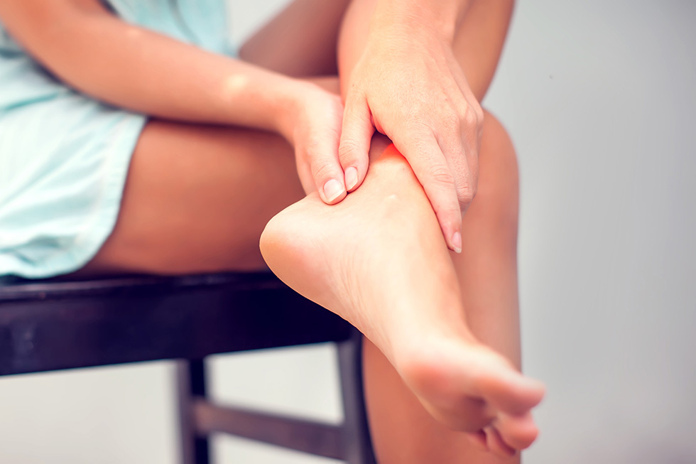 We provide comprehensive, conservative care for a wide variety of foot and ankle conditions.
 We offer same- and next-day care to patients with acute injuries.
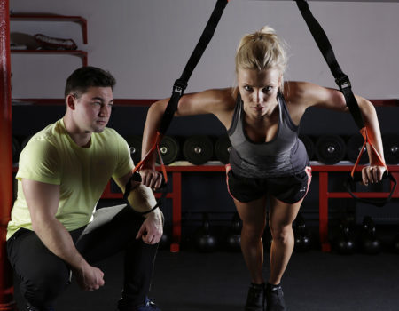 Get back in the game with help from our sports medicine specialists.
 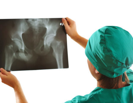 Our centers are equipped with a state-of-the-art digital X-ray machine.