


Tibiotalocalcaneal and Pantalar Arthrodesis Foot and Ankle Clinics
Aug 21, 2019George Edward Quill, M.D. (Retired 2023)
Since the report by Albert in 1882, arthrodesis has been an accepted treatment for painful arthritis of the ankle and subtalar joints.3For the purpose of this and other orthopaedic articles, surgical fusion of the ankle (tibiotalar) and subtalar (talocalcaneal) joints at the same operative sitting will be termed tibiotalocalcaneal arthrodesis. Fusion of the talus to all the bones articulating with it (distal tibia, calcaneus, tarsonavicular, and cuboid) is termed pantalar arthrodesis.(13, 20) Numerous techniques exist describing fusion approaches and implants, all having in common a similar goal: a solid, pain free arthrodesis in a biomechanically stable and functional position. (1-31)
The indications for tibiotalocalcaneal arthrodesis include avascular necrosis of the talus or a failed total ankle arthroplasty with subtalar intrusion.14,16,20,21 The failed ankle fusion with insufficient talar body, as well as patients with rheumatoid or osteoarthritic involvement of these joints are also candidates for tibiotalocalcaneal fusion.8,16,23,25,30 Other indications include the sequelae of trauma and the severe deformity of untreated clubfoot and neuromuscular disease. (5,7,20,23,24) Patients with Charcot arthropathy, skeletal defects after tumor reconstruction, pseudarthrosis, as well as fixed and various flail deformities about the hindfoot and ankle are also candidates for tibiotalocalcaneal and, in certain instances, pantalar arthrodesis. (4, 9, 18, 20,22,27,28) When there is significant instability, subluxation or arthritis involving not only the ankle and hindfoot, but also the transverse tarsal joints, then coupling the midfoot to the hindfoot by fusing the talonavicular, calcaneocuboid and calcaneonavicular articulations is indicated. (27, 29, 30)
Contraindications for tibiotalocalcaneal and pantalar arthrodesis include a dysvascular extremity or one that has a severe active infection.10, 15 Tibiotalocalcaneal arthrodesis employing medullary fixation inserted through the sole of the foot would be contraindicated in the patient with an insufficient plantar weight bearing surface for padding. Moderately severe and fixed deformity of the ankle, hindfoot and distal tibia are relative contraindications for closed nailing and arthrodesis because of the difficulty encountered in obtaining a collinear reduction of the tibia, talus, and calcaneus by the closed method. The most optimal techniques for achieving tibiotalocalcaneal and pantalar arthrodesis are described later in this paper.
The purpose of this article is to provide a relevant history of the methods and results employing tibiotalocalcaneal and pantalar arthrodesis. This author will also attempt to integrate our current knowledge and understanding of both clinical and research experience with these fusion techniques. The results and personal experience gained by the author in his own clinical series of more than 40 patients undergoing similar procedures will be presented.
History of Tibiotalocalcaneal and Pantalar Arthrodesis
A paper read at the 1922 American Orthopaedic Association in Washington, D.C., by Arthur Steindler reviewed methods of fusion about the talus published before 1922. Also, Steindler’s technique for treating the flail ankle with pan-astragaloid arthrodesis by way of denuding articular cartilage was therein described. (27)
In 1936, W.R. Hamsa, adding follow-up data from his own experience and from Dr. Steindler’s service in Iowa City, offered data on 85 patients followed after pan-astragaloid arthrodesis for neuromuscular imbalance or flail feet and ankles.12 Manuel Bastos Ansart of Barcelona, Spain, also published on results of pan-arthrodesis for paralytic flail foot in 1951.4
Liebolt recommended in 1938 that the “pantalar” arthrodesis be accomplished in two stages because of prolonged operating time and the difficulty in obtaining correction of all deformities simultaneously. He recommended doing the subtalar arthrodesis first, later fusing the ankle when it is easier to obtain the desired final degree of equinus/dorsiflexion.20 A one-stage operation for pantalar arthrodesis was published by Hunt, et al, in 1954. In this operative technique, the authors used an anterolateral approach to disarticulate and remove the talus from the hindfoot. In this technique, the talus is then laid on a block and peeled of its cartilage, cortex and ligaments. Its approximate size and shape are reportedly retained, the articular cartilage of the tibia, fibula, calcaneus and navicular is then roughened with a small gouge, and the denuded talus is replaced in its bed. This report does not include any data regarding the incidence of talar AVN following such a procedure.13 In this technique, as well as most others done for the residuals of anterior poliomyelitis that was often associated with knee extensor weakness, it was recommended that the ankle and hindfoot be fused in 8 to 10 degrees of plantar flexion to help stabilize the knee in slight recurvatum during stance.1, 4, 13, 18,20,27,28
Blair described in 1943 a sliding tibial graft that was fixed to the talar head and neck to obtain tibiotalar fusion in cases of comminuted fractures and fracture/dislocations of the body of the astragalus, resulting in either avascular necrosis or significant collapse.7 Marek and Schein reported on aseptic necrosis of the astragalus following arthrodesing procedures of the tarsus in October of 1945. These authors concluded that aseptic necrosis of the body of the astragalus was not an infrequent complication after arthrodesing procedures done to correct severe deformities of the foot. They insisted on safeguarding necrotic bone from weight bearing until it becomes revascularized postoperatively.21
Clinical evaluations after pantalar arthrodesis, some with very long-term follow-up for up to 33 years, are offered by Barrett, et al, Waugh, et al, Vahvanen, and Papa and Myerson.5,22,23,30,31 These authors conclude that extended arthrodesis of the ankle and hindfoot is a technically demanding and complicated procedure. Current scientific opinion regards pantalar and tibiotalocalcaneal arthrodesis as a salvage operation capable of producing satisfactory results when applied by experienced surgeons for the right indications. These procedures usually provide a very reasonable alternative to what otherwise would be severely disabling conditions or even amputation if left untreated. Many authors have concluded that various hindfoot and ankle arthrodeses can result in a high percent of satisfactory results, but that talectomy often did not.
Fusion of the tibia to the talus and the talus to the os calcis has been performed using posterior extra-articular methods employing a technique described by Staples.26 Intramedullary fixation for tibiotalocalcaneal arthrodesis is not a new concept. In 1906 Lexer described a technique he called “Bolzungs arthrodese”, which consisted of placing a boiled corpse bone pin through a hole in the calcaneus, talus, and tibia.19 Lexer’s method was later employed by others using an ivory peg instead of cadaveric bone. In 1915, Albee used the fibula as a vertical spike to perform an ankle and subtalar fusion.2
Adams performed a tibiocalcaneal arthrodesis in 1948 with a long three flanged nail driven from the os calcis into the tibial shaft.1Leikkonen (1950) similarly placed a metal spike from the calcaneus through the talus and tibia for ten days after performing a primary fusion.18 Küntscher recommended placing an intramedullary nail through the foot into the femur for simultaneously ankylosing the ankle and knee joints.17 Bingold (1956) placed a fibular graft through a hole made in the calcaneus, talus, and tibia by a cannulated drill over a guide wire.6 Küntscher again addressed the subject in 1967 by describing a conical nail placed from the calcaneus to the tibia for arthrodesis of the ankle and subtalar joints.17
In attempting ankle arthrodesis in the presence of ongoing sepsis, Cierny (1989) placed an intramedullary nail from the knee into the calcaneus for large central defects not amenable to external fixation.10 Two vertical Steinmann pins were placed by Carrier in 1990 for stabilization after preparing the ankle and subtalar joints.8 In 1990, Stone used a trifin nail from the calcaneus to the tibia as a primary procedure similar to Adams’ salvage technique.28 Casadei used Grosse-Kempf nails and Küntscher rods to perform ankle and subtalar arthrodesis for stability after tumor resection and reconstruction.9 In 1994, Kile and Johnson reported on a series of tibiocalcaneal arthrodeses using a custom, straight nail through a plantar incision.15
This author will include in this chapter his own clinical series of 40 patients with greater than two years’ follow-up who underwent tibiotalocalcaneal arthrodesis using an intramedullary supracondylar nail inserted through the plantar aspect of the foot and interlocked with transverse screws proximal and distal to the ankle.
Tibiotalocalcaneal Arthrodesis
The goals of ankle arthrodesis are the relief of pain and deformity and the development of a solid fusion. Numerous techniques and instrumentation devices have been described to achieve these goals. For isolated tibiotalar arthrodesis in which the surgeon wishes to leave the subtalar joint free, the author prefers a transfibular open technique employing cannulated screws for fixation. This technique has been well described, and it has almost been exhaustively studied biomechanically.1
For patients who have disabling arthritis, infection or deformity of not only the tibiotalar, but also the talocalcaneal joint, however, the surgeon must also fuse the subtalar joint along with the ankle. This can also be accomplished with a cannulated screw technique.23 Success has been achieved with sliding fibular struts anchored to the tibia, talus and calcaneus. Plates applied laterally to the tibia, talus and calcaneus have also been employed for fixation and maintenance of the desired position of arthrodesis.
The author believes, however, that intramedullary nailing is a solid method of fixation for achieving tibiotalocalcaneal arthrodesis. A medullary nail inserted through the plantar aspect of the foot can afford excellent stability and satisfactory position and alignment. In the author’s experience, patients undergoing intramedullary fixation for tibiotalocalcaneal arthrodesis need not be casted as long, nor have their activities been restricted as severely as patients undergoing similar procedures employing other methods of fixation. Furthermore, the neuropathic patient, even in the presence of an acute Charcot process about the hindfoot and ankle can quite readily be managed with medullary fixation with very low complication rate when undergoing ankle and hindfoot arthrodesis.
In a German text entitled “The Practice of Intramedullary Nailing”, published in the 1950’s and 1960’s, Gerhardt Küntscher described a method of combined arthrodesis of the ankle and subtalar joints. He employed a technique of closed medullary nailing with a conical nail inserted over a guide pin through the sole of the foot. He felt that extensive destruction of the joint and nailing should be done simultaneously. He used a 12 to 14 millimeter nail to achieve an interference fit, and no locking of the nail was done. Patients were kept at bed rest for three weeks and then ambulated in plaster.17
In 1979, Tomeno presented 45 pan-arthrodeses using various fixation techniques, but with a fairly high infection rate. He had an 80 percent consolidation rate.29 Russotti and Johnson reported in 1988 on 21 tibiotalocalcaneal arthrodeses employing Steinmann pins and external fixation. They had radiographic union in 85 percent with satisfactory results in 75 percent employing a posterior Achilles splitting approach.25 Papa and Myerson published a series of 21 pantalar and tibiotalocalcaneal arthrodeses for osteoarthrosis.23 They achieved an 86 percent fusion rate using a transfibular approach with cannulated screws. These authors also had two patients who underwent tibiotalocalcaneal arthrodesis in a neuropathic fusion series. Both of these patients fused in under five months using a cannulated screw technique.22
Drs. Johnson and Gehrke returned to using an IM nail for tibiotalocalcaneal fusion in a presentation given at the 1993 Summer Meeting of the American Orthopaedic Foot and Ankle Society.14 This report was later published by Kile, et al.15 They positioned the patient prone and used an Achilles splitting approach, but they claimed initially that the technology involved in using the nail was more involved than other established techniques, and that the long-term results were not known as yet.
This author prefers to perform tibiotalocalcaneal arthrodesis using an intramedullary supracondylar nail (Figure 1). This nail is placed percutaneously and combined with arthrotomy and debridement of the ankle and subtalar joints. The nail is placed transcalcaneally through the talus into the tibia and locked proximal and distal to these joints with bicortical screws.
The indications for a tibiotalocalcaneal arthrodesis have been given previously in this chapter.
The intramedullary nail employed by the author in a series of 40 tibiotalocalcaneal arthrodeses is a fully cannulated, stainless steel closed section nail with an 11, 12 or 13 millimeter outer diameter in lengths of 15 centimeters, 17 centimeters, 20 centimeters, or 25 centimeters. The first two, distal holes are located 15 millimeters and 30 millimeters from the driving end for good calcaneal and talar fixation. A third distal screw is also present and can be used to gain purchase of the talus or the supramalleolar distal tibia. The transfixing holes are locked with 5 millimeter fully-threaded screws, and numerous interlocking holes are available proximal to the ankle for proximal fixation.
The nail employed in this series has an 8 degree bow in the parasagittal plane. Taking advantage of this bow and directing the bow’s apex posteriorly, the surgeon can employ a calcaneal insertion point anterior to the weight-bearing surface of the os calcis.
Surgical Technique: The patient is positioned supine on a radiolucent operating table with a well-padded bump under the ipsilateral buttock in order to internally rotate the involved extremity. We place another bump under the heel so that cross-table fluoroscopic imaging is facilitated. General or spinal anesthesia is usually used, and a thigh tourniquet greatly facilitates the plantar dissection. Intraoperative C-arm fluoroscopy is used as indicated.
An anteromedial or anterolateral ankle arthrotomy is used in order to correct what deformity may be present across the tibiotalar joint and to prepare the joint surface by removing what is left of the articular cartilage. Using an anterolateral approach the surgeon can quite readily extend the incision to the subtalar joint, also exposing and debriding this joint before medullary fixation. These arthrotomies also allow the surgeon a site for inserting bone graft if he feels it is necessary.
In cases of inflammatory arthritis such as rheumatoid disease, subtalar arthrotomy may not be necessary. The surgeon may want to consider a fibular osteotomy at the time of the hindfoot fusion if there is significant varus deformity or loss of tibial length relative to the fibula. If the fibular osteotomy is not done in these cases, then the fibula may actually hold the tibiotalar and talocalcaneal arthrodesis sites distracted postoperatively.
A longitudinal plantar approach is used, placing the incision slightly lateral to the midline, especially in the patient with significant valgus of the hindfoot and ankle. After the incision is made, blunt dissection is taken down to the plantar fascia, which is split longitudinally. The intrinsic muscles are swept medially or laterally, and the neurovascular bundle is identified at the medial portion of the wound. A sharp awl is used to make a plantar calcaneal corticotomy. The position of the awl can be checked intraoperatively with fluoroscopy. If the awl cannot be readily passed across both joint surfaces, the subtalar and tibiotalar articular surfaces may be crossed with the use of a cannulated drill over its threaded guide wire. This allows access through the calcaneus and talus to the medullary canal of the distal tibia.
This procedure opens up the tibiotalocalcaneal canal enough that a spade-tipped guide wire can be passed into the tibial medullary canal more readily. A series of progressively larger flexible reamers are passed over the guide wire and are used to prepare and enlarge the tibiotalocalcaneal canal, reaming a full to 1 millimeter larger than the anticipated nail’s outside diameter.
The nail is attached to its alignment guide after noting the appropriate direction of the nail’s 8 degree bow. Also before inserting the nail into the foot, the surgeon will note which holes are to be used for interlocking once the nail is inserted. The nail is slightly internally rotated so that when the screws are inserted from lateral to medial they will pass into the tibia, clearing the fibula.
The nail is quite readily inserted manually without need of the guide wire and then impacted. The distal aspect of the nail is countersunk within the os calcis, and usually the distal two or three screws are inserted for interlocking first. This allows good purchase of the foot by the nail and distal screws, and then further impaction can be done, giving compression across the tibiotalar arthrodesis site before the nail is interlocked in the tibia. The interlocking screws are inserted from lateral to medial using the interlocking alignment device and, when necessary, intraoperative fluoroscopy. This author rarely uses screws longer than 25 millimeters to gain bicortical purchase of the tibia. Locking screw lengths of 35 to 45 millimeters may be required to gain adequate purchase of the talus and calcaneus (Figures 2a and 2b).
Clinical Results: Forty patients (25 male and 15 female) with a mean age of 47.6 years (range = 14 to 82 years) underwent tibiotalocalcaneal arthrodesis with intramedullary fixation for diagnoses of osteoarthrosis (25 patients), rheumatoid arthritis (6 patients), pseudarthrosis after failed prior attempts at ankle fusion (3 patients), Charcot arthropathy (4 patients), destructive distal tibial giant cell tumor (1 patient), and rigid talipes equinus resulting from paraplegia (1 patient). With a mean follow-up interval of 30 months (range = 18 to 60 months), a 90 percent union rate was achieved at a mean time to union of 14 weeks.
Complications related to the nail included four instances of painful retained or fractured interlocking screws, four cases of nonunion, one of which was asymptomatic, and a single case of a 20 degree valgus malunion. One ulceration occurred in a neuropathic patient over a fractured interlocking screw, which was subsequently removed. This same patient had posterior tibial nerve entrapment due to that screw prior to its removal. The overall nonunion rate was 4 patients out of 40 or 10 percent. Only 3 of these 40 patients, or 8 percent, had a symptomatic nonunion. These 3 patients have subsequently been revised successfully.
The patients who underwent this procedure for the sequelae of post-traumatic osteoarthrosis had undergone a mean of five prior procedures each before coming to their index tibiotalocalcaneal arthrodesis with medullary fixation. The one patient who had had a giant cell tumor resected had six prior procedures, one of which was an unsuccessful attempt at ankle arthrodesis. This was successfully salvaged with tibiotalocalcaneal arthrodesis by the technique described here.
The results of this series would indicate that intramedullary nailing is a solid method of fixation for tibiotalocalcaneal arthrodesis in appropriately selected patients. We achieved an overall union rate of 90 percent at a mean time to union of 14 weeks in a large series that included patients undergoing tibiotalocalcaneal arthrodesis for a variety of indications. We had a symptomatic nonunion rate of 8 percent.
The best clinical results (100 percent union rate, 0 percent complication rate) after tibiotalocalcaneal arthrodesis with intramedullary fixation have been obtained in nonneuropathic patients undergoing their first fusion procedure for a diagnosis of primary osteoarthrosis or rheumatoid arthritis.
The significance of this report is that a method of hindfoot arthrodesis first described decades ago may be even more successfully employed today with the availability of a better fixation device (intramedullary ankle fusion nail). Fusion rates employing this nail are very high in appropriately selected patients, even in cases of salvage arthrodesis after failures of more conventional techniques of fixation.
Pantalar Arthrodesis
When one begins reading about the indications and surgical techniques employed for achieving pantalar arthrodesis, one quickly realizes three things: First these are some of the most technically demanding and involved procedures encountered in foot and ankle surgery today. Second, these procedures are usually done in the salvage setting for severely disabling or even limb threatening conditions. Third, one quickly realizes that most of the existing literature on pantalar arthrodesis is very old.
Pantalar arthrodesis proved a reliable, reproducible surgical procedure in addressing the flail foot and ankle often associated with the sequelae of poliomyelitis, paralysis, and tuberulous and bacterial infection in the earlier part of this century. Most early reports of this technique focus, therefore, on achieving a rigid stable hindfoot in satisfactory position and very few if any articles published before the 1950’s included recommendations on internal fixation.2,4,6,12,13,20,27
With the success and subsequent popularity of joint replacement surgery, spinal decompression and instrumentation, and the development of the subspecialty of orthopaedic sports medicine, much less emphasis was placed in our literature on pantalar arthrodesis.
To this day, however, patients who have post-traumatic osteoarthrosis involving both the ankle and subtalar joints still pose a difficult therapeutic challenge. High-speed motor vehicular trauma, spinal cord injury, longer life expectancy, and the popularity of sports and active lifestyle – as well as an orthopaedic appreciation of biomechanics – have again turned attention back to the foot and ankle. Modern articles deal with the topic of pantalar arthrodesis on a much more frequent basis.5, 8-11, 14-16, 22-25, 28-31
Alternatives to pantalar arthrodesis for patients with arthritis or bone deficiency of the ankle and hindfoot include Syme or below-the-knee amputation.23 We are learning much more currently about the expected functional results in patients who have an arthrodesis of the hindfoot and ankle.11 Debate continues regarding the relative attributes of a one-stage versus two-stage technique in achieving pantalar arthrodesis.4-6,9,10,13-15,21,24-27 For reasons of economics, patient desire for expedient care, and an interest to rehabilitate the patient more quickly and return to more normal lifestyle, this author prefers a one-stage procedure.
A satisfactory pantalar arthrodesis can be achieved employing the intramedullary nail for tibiotalocalcaneal arthrodesis, and in turn, linking this fused hindfoot to the midfoot with cannulated screws or bone staples (Figure 3). An alternative treatment method employs performing a triple arthrodesis of the foot in standard fashion and then linking this to the ankle in the desired position of neutral dorsiflexion/plantar flexion, 3 to 5 degrees of hindfoot valgus, and external rotation symmetric with that of the uninvolved contralateral side. Every effort should be made to keep the foot plantigrade.11
The first and fifth metatarsal heads must strike the ground at the same time. If the ankle and hindfoot are fused in too much equinus, the patient will have a tremendous tendency to recurvatum at the knee and have heel-off too early in the gait cycle. These patients will often walk with the involved extremity externally rotated in an effort to more easily vault over the involved foot that is in equinus.
If the ankle and hindfoot are fused in too much dorsiflexion, the gait will seem more natural and stride length more symmetric with the other uninvolved side. However, the pressures of weight bearing at heel strike will quickly become uncomfortable, and the patient will lack satisfactory push-off.
Gellman and others have shown that the deficits in dorsiflexion and plantar flexion after isolated tibiotalar arthrodesis are 50.7 percent and 70.3 percent respectively. After tibiotalocalcaneal arthrodesis, however, the dorsiflexion and plantar flexion deficits are only 53 percent and 71.3 percent respectively. Thus, linking the calcaneus to the fused ankle does not cause an appreciable loss of dorsiflexion or plantar flexion. Inversion and eversion, however, undergo a diminution at least 40 percent greater after tibiotalocalcaneal arthrodesis than they do with ankle fusion alone.11
Pantalar arthrodesis has been noted to cause deficits in dorsiflexion and plantar flexion of 62.8 percent and 82.2 percent respectively. Inversion and eversion are reduced by 71.7 percent and 67.4 percent respectively after pantalar arthrodesis.11 These values stress the absolute importance of achieving appropriate position of the foot and ankle if the pantalar arthrodesis is to be successful.
Operative Technique: Pantalar arthrodesis is performed with the patient supine with a wellpadded bump placed underneath the ipsilateral buttock. A pneumatic tourniquet is used at the level of the thigh, and the ipsilateral iliac crest prepared, as well as the entire lower extremity from the knee distally. It is also quite helpful to note, if one can palpate through the sterile drapes or have prepped into the field, the contralateral uninvolved lower extremity to help one ascertain appropriate position and rotational alignment. The technique of Papa and Myerson is preferred.23
An extended lateral approach is made, employing an incision longitudinally oriented over the distal fibula and curving anteriorly over the sinus tarsi. One must take care to avoid the cuticular branches of the superficial peroneal nerve and the sural nerve. A distal fibular ostectomy is performed with an oscillating saw at a level just proximal to the tibiotalar joint. In this fashion wide exposure of the ankle and hindfoot is obtained, and yet the proximal fibula is still well anchored to the tibia by the syndesmotic ligaments. The cut may be bevelled from proximal lateral to distal medial and the distal portion of the fibula saved and used as autogenous bone graft in piecemeal fashion.
A second anteromedial incision can be made midway between the tibialis anterior tendon and the medial malleolus, protecting the greater saphenous vein and its cuticular nerve. This incision can be extended distally and posteriorly if necessary for greater exposure. The second incision can be used to either resect the medial malleolus if it is unduly prominent, or preferably, leave the medial malleolus in place and denude the articular cartilage from this corner of the ankle joint.
If great deformity is not present through the ankle joint, one can use a sharp bone chisel, and in a congruous fashion, remove the articular cartilage and hard subchondral bone, taking appropriate resections of bone as necessary to make the foot plantigrade. Alternatively, as described by Papa and Myerson, an oscillating saw held perpendicular to the long axis of the tibia can be used to make planar cuts, preserving as much bone stock as possible.23 A lamina spreader is a great aid in exposing the subtalar and transverse tarsal joints. Chisels, rongeurs and curettes can be used to denude the articular cartilage from these joints and appropriate wedges of bone removed as necessary to make the foot plantigrade. Final alignment of the hindfoot in 0 to 5 degrees of valgus, 0 to 5 degrees of calcaneus, and external rotation equal to that of the contralateral extremity is desired. Normally, the longitudinal axis of the anteromedial crest of the tibia and the tibial tubercle line up with the second ray of the normal foot. In earlier literature a position of 5 to 10 degrees of equinus was recommended, but these pantalar arthrodeses were then being done for a flail foot in the presence of quadriceps weakness, in which case plantar flexion floor reaction forces causing recurvatum at the knee were desired. In the patient with normal quadriceps, 0 to 5 degrees of calcaneus is more desirable.
Fixation can be achieved with threaded 2 millimeter guide wires, facilitating 6.5 or 7 millimeter cannulated screw fixation. An autogenous graft harvested from either the distal fibula and/or the anterior iliac crest can be tamped into available spaces to insure mechanical support and quicker healing.
The author finds it easiest to fix first from dorsal to plantar with guide pin and cannulated screw the talocalcaneal joint. Next, taking care to rotate the foot appropriately through the transverse tarsal joints, a proximal to distal calcaneocuboid screw and a distal to proximal talonavicular screw can be inserted over guide wires.
Making sure that the talocalcaneal screw is anterior and inferior to the lateral process of the talus, a guide wire is next inserted from just anterior to the lateral process of the talus in a distal to proximal and lateral to medial direction across the tibiotalar joint. On the lateral view, this wire and subsequent cannulated screw would be passed in the anterior body of the talus, exiting the medial tibia at the supramalleolar level. I have found it easier to insert this screw, however, over this guide wire from proximal, medial to distal lateral, gaining better purchase in the talus with the threads and countersinking the head of the screw in the medial cortex of the metaphyseal tibia.
The last screw can be placed anterolateral on the distal tibia to posteromedial in the posterior body of the talus. On the lateral x-ray this screw appears to be parallel to the first tibiotalar screw, and on the AP film appears to cross the medial tibial talar screw at nearly a right angle.
After further irrigation and bone grafting, the wounds can be closed in layers over suction drainage tubes, and a bulky dressing reinforced with plaster splints is applied. The drains can be removed 24 to 48 hours postoperatively with the patient being discharged from the hospital in the routine case within 48 hours from surgery.
At approximately two weeks postoperatively, if there are no special circumstances, the operative bandages and sutures are removed, and a well-padded, well-molded short leg cast is applied. The patients are routinely kept nonweight bearing for six weeks, followed by weight bearing to tolerance the subsequent six weeks. Clinical and radiographic correlation are used to modify this recommended postoperative regimen. Shoe lifts may be necessary to compensate any limb length discrepancy caused by the shortening inherent in this procedure. Ideally the operated extremity should be approximately one-half to one centimeter shorter than the contralateral uninvolved limb to allow clearance in the swing phase of the gait cycle.
Most patients are able to wear regular athletic or walking shoes after a period of adjustment and experimentation upon removing the final cast. Some patients, however, will benefit greatly from the application of a rocker sole or single axis cushioned heel to the plantar aspect of the shoe on the involved side.
Complications after pantalar arthrodesis include superficial and/or deep wound infection, wound slough, malunion and nonunion. Hardware failure or prominence can be remedied with subsequent removal after radiographic union. Cuticular neuromas have been reported, and prevention is the key to minimizing symptoms here. Patients with normal plantar sensation and arthrodesis in the appropriate position should have minimal incidence of ulceration.
Avascular necrosis was reported after pantalar arthrodesis primarily when talectomy and reinsertion of the talus was employed in earlier reports.13, 21
The ankle is the most frequently reported site of pseudarthrosis after a pantalar arthrodesis, followed by the talonavicular joint. The more rigid the fixation and the more the surgeon respects the local vascular anatomy, the less the likelihood of nonunion developing.
Malunion is a fairly frequent complication of pantalar arthrodesis because of the great technical demands of performing this procedure. A precise determination of appropriate final alignment is the key. Intra-operative radiographs should be employed in almost every case. Hindfoot valgus seems to be better tolerated than varus, and if the surgeon had to error, he should do so to the side of valgus. Avoidance of internal rotation and the use of mild posterior subluxation of the talus under the tibia can enhance postoperative gait.
In conclusion, the reader will appreciate that tibiotalocalcaneal and extended pantalar arthrodesis are quite demanding, but useful procedures that may be employed for a variety of indications. These procedures should be viewed as salvage techniques; to be employed for what otherwise would be very disabling or even limb threatening situations for patients with osteoarthrosis, rheumatoid arthritis, neuropathic joint destruction, and paralytic or flail extremities.
Bibliography
- Adams, J.C.: Arthrodesis of the Ankle Joint. Experiences With the Transfibular Approach. J. Bone and Joint Surg., 30B:506-511, 1948.
- Albee, F.H.: Bone-Graft Surgery, Philadelphia and London: W.B. Saunders, p. 335, 1915.
- Albert, Eduard: Einige Fälle von künstlicher Ankzlosen bildung an paralytischen Gleidmassen. Weiner Medizinische Press, 1882.
- Ansart, M.B.: Pan-Arthrodesis for Paralytic Flail Foot. J. Bone and Joint Surg., 33-B: 503- 507, 1951.
- Barrett, G.R., Meyer, L.C., Bray, E.W., Taylor, R.G., and Kolb, F.J.: Pantalar Arthodesis: A Long-Term Follow-up. Foot and Ankle, Volume 1 #5:279-283, 1981.
- Bingold, A.C.: Ankle and Subtalar Fusion by a Transarticular Graft. J. Bone and Joint Surg., 38B:862- 870, 1956.
- Blair, H.C.: Comminuted Fractures and Fracture Dislocations of the Body of the Astragalus: Operative Treatment. American J. of Surgery, Vol. LIX (1):37-43, 1943.
- Carrier, D.A., and Harris, C.M.: Ankle Arthrodesis with Vertical Steinmann’s Pins in Rheumatoid Arthritis, CORR 268:10-14, 1991.
- Casadri, R., Ruggieri, P., Giuseppe, T., Biagini, R., and Mercuri, M.: Ankle Resection Arthrodesis in Patients with Bone Tumors. Foot and Ankle International, 15:242-249, 1994.
- Cierny, G., Cook, G., and Mader, J.: Ankle Arthrodesis in the Presence of Ongoing Sepsis. Orthop. Clinics of North America, 20:709-721, 1989.
- Gellman, H., Lenihan, M., Halikis, N., Bott, M., Giordani, M., and Perry, J.: Selective Tarsal Arthrodesis: An In Vitro Analysis of the Effect on Foot Motion. Foot and Ankle, Vol. 8(3):127- 133, 1987.
- Hamsa, W.R.: Panastragaloid Arthrodesis. J. Bone and Joint Surg., 18:732-736, 1936.
- Hunt, W.S., and Thompson, H.A.: Pantalar Arthrodesis: A One-Stage Operation. J. Bone and Joint Surg., 36-A: 349-363, 1954.
- Johnson, K.A., and Gehrke: Proceedings of the American Foot and Ankle Society 1993 Summer Meeting, Intramedullary Fixation for Ankle Arthrodesis.
- Kile, T.A., Donnelly, R.E., Gehrke, J.C., Werner, M.E., and Johnson, K.A.: Tibiocalcaneal Arthrodesis with an Intramedullary Device. Foot and Ankle International 15:669-673, 1994.
- Kitaoka, H.B., and Romness, D.W.: Arthrodesis for Failed Ankle Arthroplasty. Journal of Arthroplasty, Vol. 7 #3:722-784, 1992.
- Küntscher, G.: Combined Arthrodesis of the Ankle and Sub-talar Joints, in Practice of Intramedullary Nailing. Charles C. Thomas: 207-209, 1967.
- Leikkonen, O.: Astragalectomy as an Ankle-Stabilizing Operation in Infantile Paralysis Sequelae. Acta Chirurgica Scandinavica Suplementum 152:13-148, 1948.
- Lexer, E.: Die Verwedung der freien Knochenplastik nebst Versuchen über Gelenkversteifung und Gelenktransplanten. Langenbecks Archive für Klin. Chirung. 86:938, 1906.
- Liebolt, F.L.: Pantalar Arthrodesis in Poliomyelitis. Surgery 6:31-34, 1939.
- Marek, F.M., and Schein, A.J.: Aseptic Necrosis of the Astragalus Following Arthrodesing Procedures of the Talus. J. Bone and Joint Surg., Vol. 27(4):587-594, 1945.
- Papa, J., Myerson, M., and Girard, P.: Salvage, With Arthrodesis, in Intractable Diabetic Neuropathic Arthropathy of the Foot and Ankle. J. of Bone and Joint Surg., Vol. 75 #7:1056- 1066, 1993.
- Papa, J.A., and Myerson, M.S.: Pantalar and Tibiotalocalcaneal Arthrodesis for PostTraumatic Osteoarthrosis of the Ankle and Hindfoot. J. of Bone and Joint Surg., Vol. 74-A #7:1042-1049, 1992.
- Reckling, F.W.: Early Tibiocalcaneal Fusion in the Treatment of Severe Injuries of the Talus. The Journal of Trauma, Vol. 12, No.1:390-396, 1972.
- Russotti, G.M., Johnson, K.A., and Cass, J.R.: Tibiotalocalcaneal Arthrodesis for Arthritis and Deformity of the Hind Part of the Foot. J. of Bone and Joint Surgery. Vol. 70-A #9:1304- 1307, 1988.
- Staples, O.S.: Posterior Arthrodesis of the Ankle and Subtalar Joints. J. Bone and Joint Surg., 38-A (1):50-58, 1956.
- Steindler, A.: The Treatment of the Flail Ankle; Panastragaloid Arthrodesis. J. Bone and Joint Surg., 5:284-294, 1923.
- Stone, K.H., and Helal, B.: A Method of Ankle Stabilization. CORR 268:102-106, 1991.
- Tomeno, B., and Danan, J.P.: Pan Arthrodesis of the Rear of the Foot. Revue de Chirurgie Ortopedique, Vol. 65:433-439, 1979.
- Vahvanen, V.: Arthrodesis of the TC or Pantalar Joints in Rheumatoid Arthritis. Acta Orthopaedics Scandinavia, Vol. 40:642-652, 1969.
- Waugh, T.R., Wagner, J., and Stinchfield, F.E.: An Evaluation of Pantalar Arthrodesis. J. Bone and Joint Surg., Vol. 47-A (7):1315-1322, 1965.
Legends for Figures
- Figure 1: An example of the intramedullary nail used for fixation of tibiotalocalcaneal arthrodeses.
- Figure 2a: Preoperative radiograph of a 41-year-old basketball coach with advanced osteoarthrosis of the ankle and hindfoot.
- Figure 2b: Postoperative radiograph obtained six weeks after the patient pictured in Figure 2a underwent tibiotalocalcaneal arthrodesis with medullary nail fixation and autogenous bone graft.
- Figure 3: Postoperative radiograph of a 60-year-old woman with rheumatoid arthritis who underwent a successful pantalar arthrodesis employing the intramedullary ankle fusion nail (see text).


 Our patients can receive MRI imaging onsite at both our Louisville and New Albany Clinics.
 Providing the latest advances in orthopedic surgery is our specialty.
 We take a unique, multidisciplinary approach to pain management.
 Our physical therapists use advanced techniques to help restore strength and mobility.
 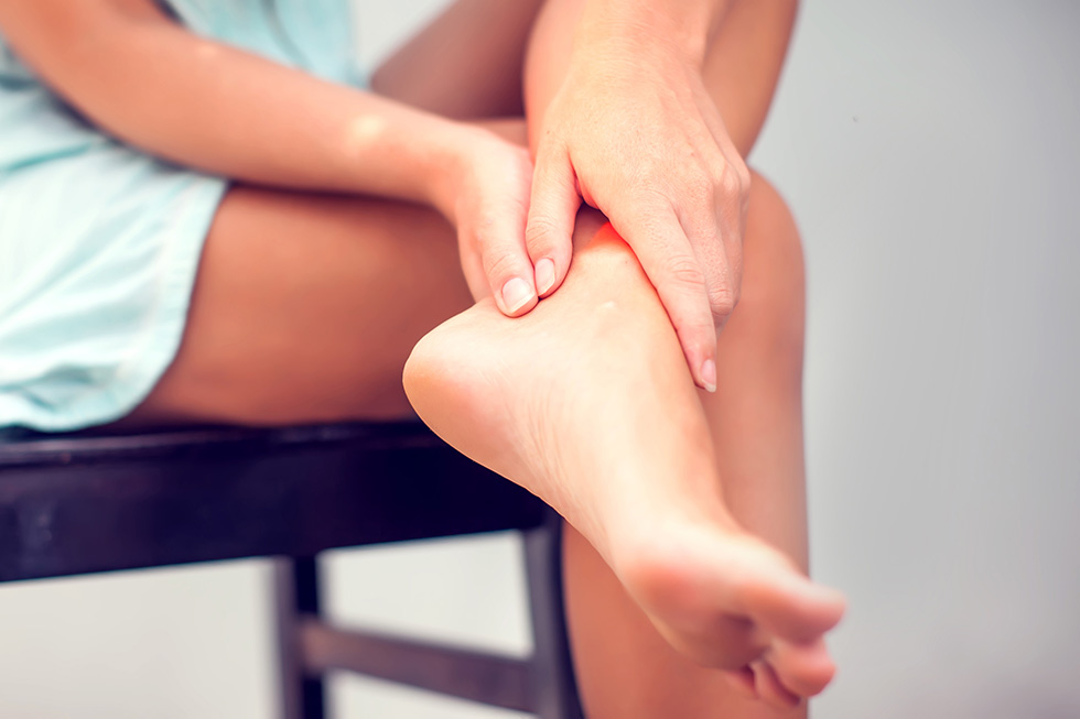 We provide comprehensive, conservative care for a wide variety of foot and ankle conditions.
 We offer same- and next-day care to patients with acute injuries.
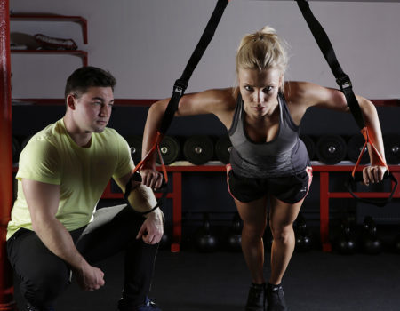 Get back in the game with help from our sports medicine specialists.
 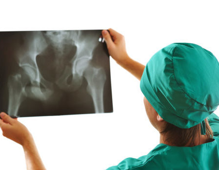 Our centers are equipped with a state-of-the-art digital X-ray machine.